The muscles at the back of your thigh are an essential component of your lower kinetic chain, providing stability and contributing to efficient locomotion. The hamstrings cross over two joints – the hip and the knee – assisting in knee flexion and hip extension. Pain and dysfunction in your hamstring muscles can affect your gait, altering your static and dynamic posture and increasing your risk of injury.
Your hamstrings are a group of three muscles at the back of your upper leg. Because the hamstrings cross over two joints, they perform dual functions – knee flexion, and assisting with hip extension. The hamstrings play an important role in the gait cycle, and they are essential for running, jumping, and lifting objects off the ground, The hamstrings directly affect pelvic alignment and joint alignment along the lower kinetic chain. Weak, tight, and/or lax hamstrings can cause back pain, knee pain, hip pain, pelvic misalignment, and other issues.
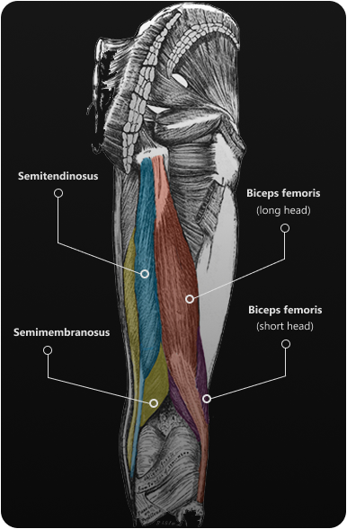
The hamstring muscle group includes:
Biceps femoris
– a muscle with a long and a short head that runs down and across your posterior thigh. The two heads share a common tendon at the back of your knee that attaches to the head of the fibula of your lower leg. The biceps femoris long head originates from the ischial tuberosity of your pelvis. The short head originates at the posterior surface of your femur, or thigh bone.
Semitendinosus
– a tendinous muscle that runs the length of your thigh next to the biceps femoris. Like the biceps femoris, the semitendinosus originates from the ischial tuberosity of your pelvis, but the distal end attaches to the medial surface of your tibia.
Semimembranosus
– a broad flat muscle located beneath the semitendinosus. It also originates from the ischial tuberosity, but slightly farther up, and attaches to the medial tibial condyle.
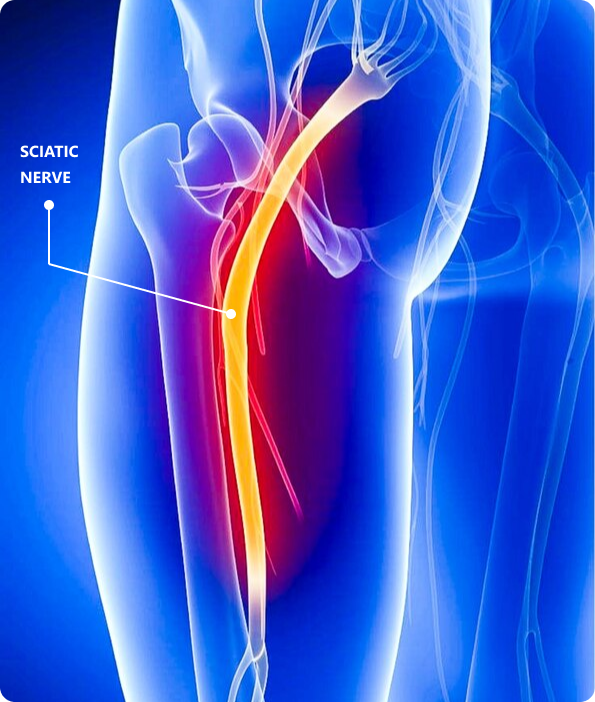
The hamstring muscles are innervated by the sciatic nerve that descends from your lumbar spine and runs down the back of your upper leg. Compression or entrapment of the sciatic nerve can seriously impede hamstring function.
or

Clinical director & DC RMSK
Verified Expert Profiles
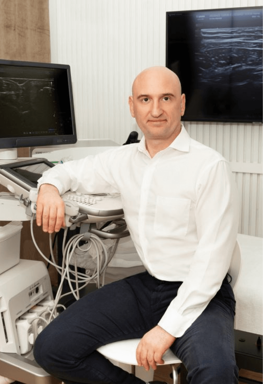
With over 25 years of experience treating musculoskeletal disorders, Dr. Kalika has formulated his own unique approach to hamstring diagnosis and treatment. As an expert in diagnostic ultrasonography, he has published multiple scientific publications that have helped to take diagnostic medicine to the next level. He has worked with some of the world’s leading radiologists in the field of musculoskeletal ultrasonography.
Dr. Kalika has helped hundreds of patients to rehabilitate hamstring injuries, restore function, and return to sports and physical activity. Dr. Kalika’s expertise in musculoskeletal ultrasonography, shockwave therapy, regenerative medicine and 3D gait analysis makes him one of the most sought-after specialists in NYC for hamstring pain and dysfunction.
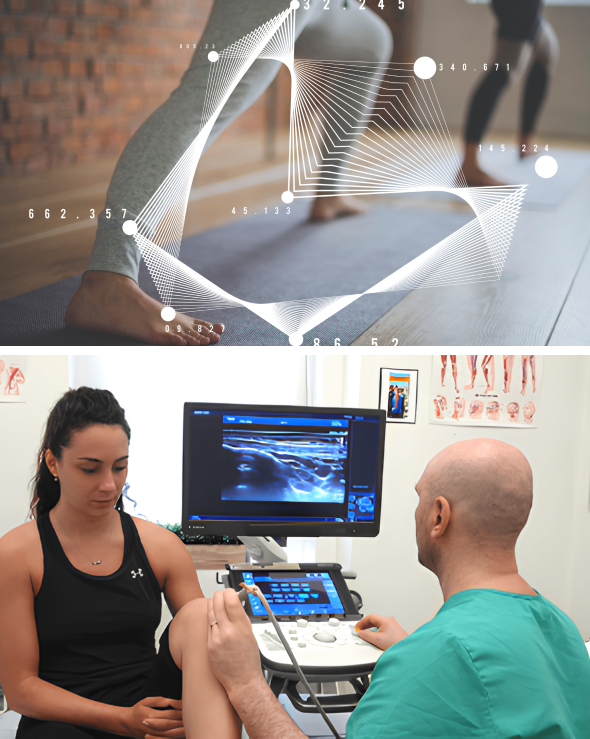
NYDNRehab features research-grade ultrasonography with the highest resolution available in New York City. Our equipment gives us capabilities for sonoelastography to test for tendon stiffness, and superior microvascular imaging (SMI) to assess inflammation and detect vascular neogenesis.
NYDNRehab is among the first sports medicine clinics to feature dynamic ultrasonography using the USONO ProbeFix device. ProbeFix attaches directly to the athlete, allowing us to visualize damaged tissues during sport-specific actions. ProbeFix can even be synced with motion capture cameras to produce 3D images of muscles, fascia, bones and joints during physical activity. This game-changing technology gives us a huge advantage for diagnosing and treating hamstring injuries.
Click here to learn more about our diagnostic approach to hamstring injuries…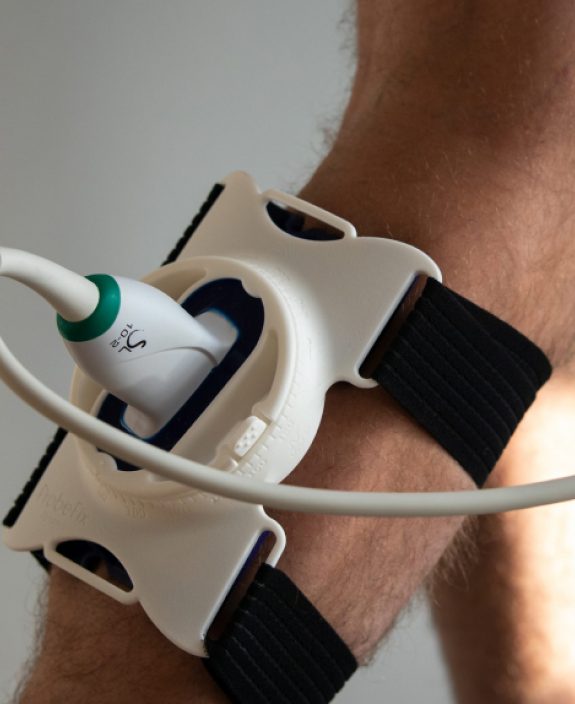
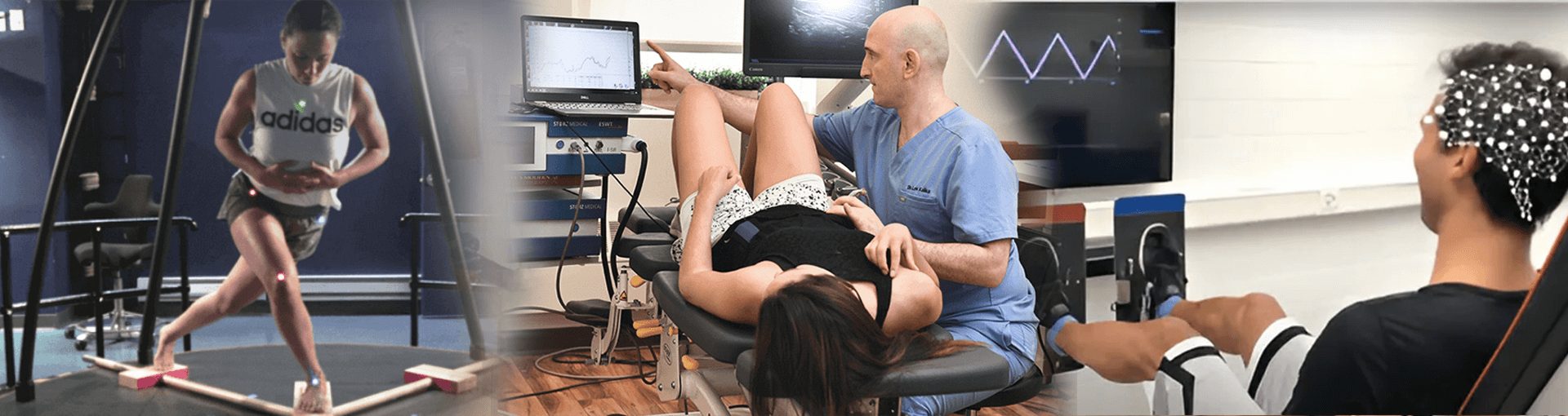
NYDNRehab is the only clinic in New York with a fully equipped motion and gait analysis laboratory. Our motion and gait analysis technology and proprietary software let us measure and quantify hamstring function in real time.
For sport-specific injuries, we use the latest technologies to analyze movement mechanics and identify motor deficiencies. By eliminating mechanical and training errors, athletes and physically active patients are able to dramatically improve their performance while reducing their risk of injury.
Sports-related hamstring injuries are the most common athletic injuries, occurring frequently in sports like sprinting, soccer, baseball and American football. Many injuries occur at the musculotendinous junction (MTJ) where muscles, tendons and bones converge. In general, the closer the injury is to the MTJ, the longer it will take to heal and regain function.


Torn or damaged tissues, with no reduction in strength
Moderate damage to tissues with some loss in strength
Severe injuries with tissue ruptures or avulsions, with significant loss of strength
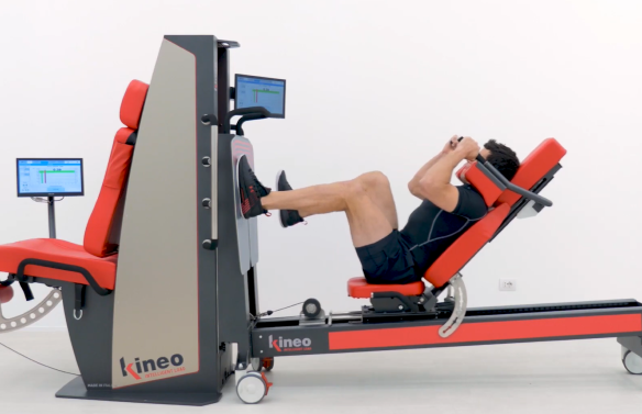
At NYDNRehab, we use the Kineo Intelligent Load system to compare strength between the injured and non-injured leg in both the concentric and eccentric phases of muscle contraction. We confirm our diagnosis with dynamic high resolution ultrasound imaging to view the structures of the hamstrings, low back, and lower kinetic chain in real time.
Ultrasound lets us travel the length of the sciatic nerve to look for damage or entrapment. It also enables sonoelastography, to gauge the relative elasticity of damaged tendons. Ultrasound imaging takes place in our clinic during your very first visit, with no waiting for lab results.
High-resolution ultrasonography not only gives us a cutting-edge diagnostic tool – it is a game-changer when it comes to hamstring treatment. Most injuries involve multiple tissue types, and each demands its own therapeutic approach. Ultrasound imaging lets us differentiate between various tissues and structures, to ensure that our needling and regenerative procedures hit their mark.
Ultrasound also provides a dynamic feedback tool for motor retraining. Patients often develop compensation patterns after a hamstring injury to off-load injured tissues, disrupting optimal muscle coordination patterns. Once the tissues have healed, it is necessary to restore neuromuscular pathways that govern motor unit firing patterns. Ultrasound gives the patient visual feedback, to help restore optimal muscle activation.
In addition, ultrasonography helps us track and confirm the effectiveness of our treatment protocols. Its capabilities for sonoelastography and superb microvascular imaging let us detect early signs of healing, and dynamic real-time imaging helps us measure the patient’s progress.
Most people take everyday mobility for granted until an injury occurs or pain sets in. Sometimes pain and reduced mobility seem to arise out of nowhere, with no apparent cause of onset. Regardless of whether your pain is caused by trauma or by something less obvious, tensegrity plays a key role.
Tensegrity refers to tensile integrity – a state where a system of individual components is held together under continuous elastic tension. In the human body, tensegrity is created by the myofascial system, the network of muscles and fascia that work together to produce, control, and guide forces, and to hold the body’s various organs and structures in place during movement.
Tensegrity can be disrupted when myofascial tissues are injured or damaged in some way. When that happens, nerves and blood vessels can become entrapped, preventing them from gliding among other structures and producing pain. At the same time, the elastic tension that governs joint alignment and controls movement becomes compromised, creating motor deficits that undermine mobility and stability.
Factors that disrupt myofascial tensegrity include:
Many doctors do not understand the crucial role of the myofascial system in preventing pain syndromes, movement disorders, and disease. In fact, most medical doctors have no idea how to correct myofascial dysfunction or even recognize it as a factor. They simply treat pain symptoms with medications and eventually recommend surgery.
At NYDNRehab, we understand that the body’s systems work together as an integrated whole, and that treating pain is not enough to eliminate its source. We use dynamic high-resolution ultrasound to explore the myofascial system in real time. Ultrasound imaging lets us visualize muscles, fascia, nerves and other structures in motion, to identify places where tensegrity has been disrupted.
Once we identify the problem, we use the most advanced therapeutic approaches to restore myofascial integrity and promote tissue healing.

Most injuries involve more than one type of tissue, and identifying and treating damaged issues prior to beginning physical therapy is key to getting fast and effective results. Failure to pre-treat your condition can completely undermine your treatment protocol, and in some cases, your condition may even worsen.
Obstacles to physical therapy success include:
At NYDNRehab, we use a broad range of regenerative technologies and integrative therapeutic approaches to resolve issues that can stand in the way of successful physical therapy. Our staff is certified in a diverse array of holistic treatment methodologies, and our one-on-one treatment sessions are personalized, based on your unique diagnostic profile.
Once we pre-treat your damaged tissues and eliminate compensation patterns, your body will be ready to begin physical therapy.
The human body is made up of integrated parts and systems, designed to work in harmony. Today’s modern reductionist approach to medical treatment fails to acknowledge the integral relationship between structure and function, focusing on the locus of pain without considering its broader implications.
Most injuries involve multiple structures and tissue types, and pain is only a symptom that signals your brain to protect and heal the damaged tissues. It is not uncommon for an injury to stop hurting while damaged structures remain dysfunctional.
At NYDNRehab, our diagnostic process takes into account the interrelated nature of the body’s structures and systems, along with the unique anatomical characteristics of the individual patient. We use dynamic high resolution ultrasound to visualize your injury in real time. Diagnostic ultrasound helps us determine whether pain and dysfunction are caused by structural (anatomical) changes, or if compensation strategies are causing structural changes. Your ultrasound exam takes place on-site, on your first visit, so we can quickly set you on the road to recovery.
The human body has its own innate healing mechanisms, but hamstring tissues sometimes need a nudge to accelerate the healing process. Regenerative technologies help to jump-start hamstring healing by stimulating tissue repair at the cellular level. Our regenerative therapies expedite recovery with minimal discomfort for the patient.
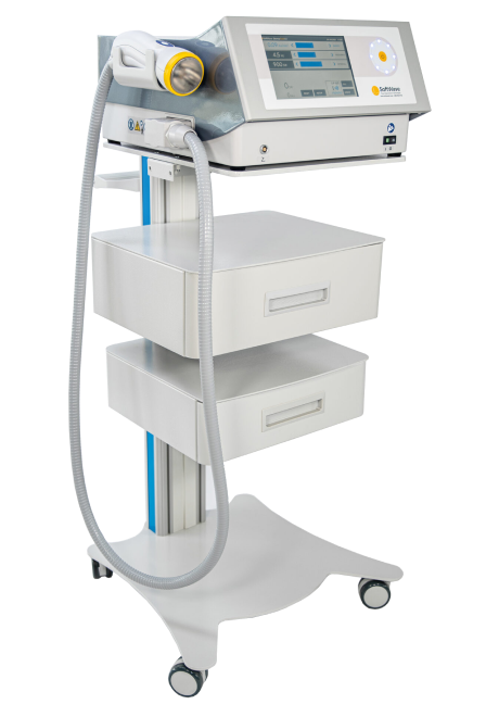
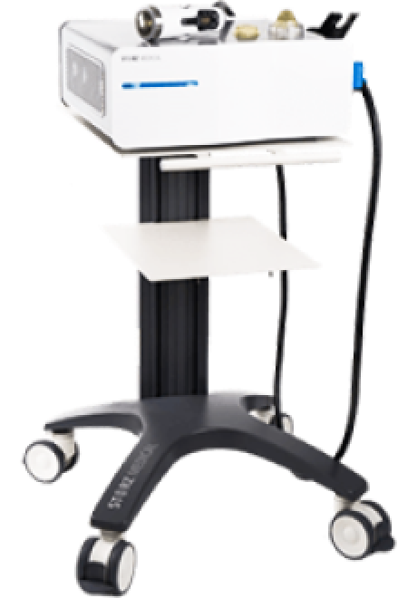
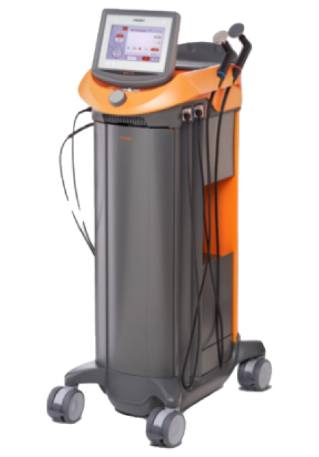
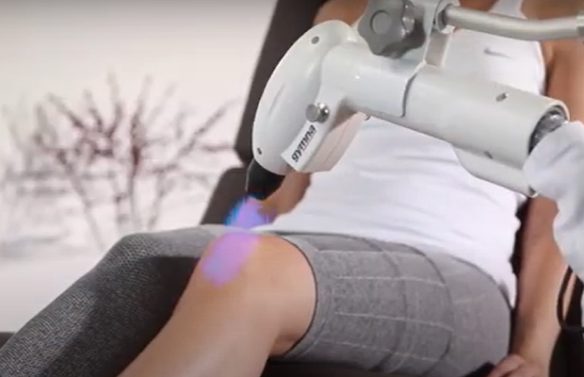
SoftWave is a groundbreaking regenerative mechanotransduction technology that accelerates tissue healing. Its patented electro-hydraulic applicator delivers high-speed soundwaves that can penetrate up to six inches in depth. SoftWave’s defocused and linear focused shockwaves recruit maximum stem cells to the treatment site to promote healing. SoftWave’s wider and deeper penetration using defocused energy is a preferred treatment option for a broad spectrum of conditions, ranging from orthopedic injuries to pelvic health. SoftWave is the only unfocused shockwave technology currently available. According to recent research, SoftWave defocused waves combined with focused and radial shockwaves have maximum regenerative potential.
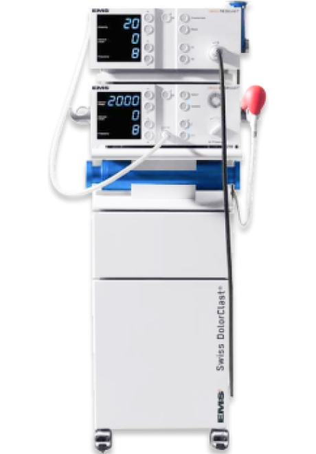
MyACT is a new type of focused shockwave technology that allows for deeper compression of the focused waves. Its higher frequency allows for precise neuro modulation under ultrasound guidance, with a special linear head for treating myofascial pain. MyACT transforms the mechanical energy of shockwaves into biochemical signals that precisely target damaged tissues. Most injuries involve more than one tissue type. When used together, our advanced shockwave technologies enable us to specifically target multiple tissue types with the most effective shockwave treatment.
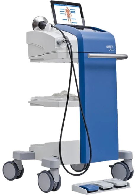
Focused ESWT is used as a regenerative treatment for damaged tendon, muscle and bone tissue. This technology produces high frequency sound waves to stimulate the body’s own reparative mechanisms. It is especially effective for chronic degenerative tendon disorders and myofascial pain syndrome.
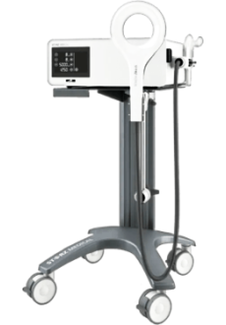
EMTT transmits high energy magnetic pulses to targeted tissues that synchronize with the body’s own magnetic fields, triggering a regenerative response. EMTT waves can penetrate deep tissues to target difficult-to-reach tendons, muscles, bones and nerves.
EPAT, sometimes called defocused shock wave therapy, is not a true shockwave. It uses mechanical pressure waves to enhance blood circulation, improving oxygen and nutrient delivery to muscle and fascia tissues, but has minimal regenerative properties.The mechanical properties of EPAT make it especially effective for fascial manipulation in combination with focused shockwaves. We combine EPAT with different types of shockwaves for holistic treatment, without additional cost to the patient.
HEIT delivers high-intensity magnetic pulses to peripheral nerve tissues, to stimulate neuroplasticity. We leverage this FDA-approved methodology to treat pain and regenerate nerve fibers, for enhanced motor control.
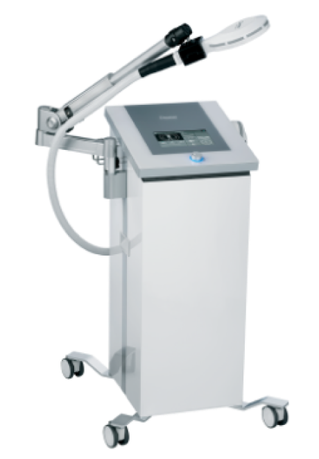
INDIBA is a form of TECAR therapy that helps to restore the ionic charge of damaged cells, for faster injury healing and rehabilitation.
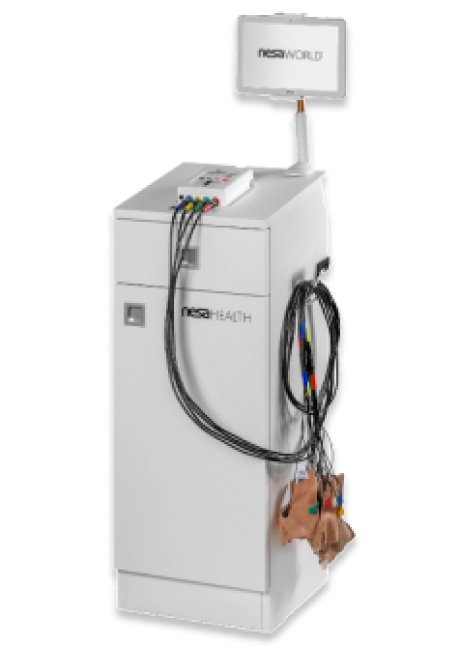
NESA generates a low-frequency electrical current of intermittent and cyclical stimuli that soothes hypersensitized nerves and restores optimal signaling between the autonomic nervous system and the brain. We leverage this FDA-approved methodology to treat pain and regenerate nerve fibers, to enhance motor control.
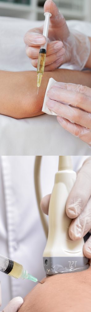
Injection therapies use orthobiologic solutions that stimulate cellular repair by either nourishing or irritating the targeted cells. Dr. Kalika teams up with an orthobiologic injection specialist to deliver ultrasound-guided needling procedures. Guidance by ultrasound ensures that the injected substances hit their mark, for maximum effectiveness.
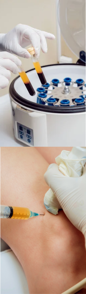
PRP therapy uses a sample of the patient’s own whole blood, which is spun in a centrifuge to extract a high concentration of platelets. When injected into damaged tissues, PRP initiates tissue repair by releasing biologically active factors such as growth factors, cytokines, lysosomes and adhesion proteins. The injected solution stimulates the synthesis of new connective tissues and blood vessels. PRP can help to jump-start tendon healing in chronic injuries and accelerate repair in acute injuries.
Alpha 2 macroglobulin (A2M) is a naturally occurring blood plasma protein that acts as a carrier for numerous proteins and growth factors. As a protease inhibitor, A2M reduces inflammation in arthritic joints and helps to deactivate a variety of proteinases that typically degrade cartilage.
Prolotherapy uses a biologically neutral solution to irritate stubborn tissues, triggering the body’s innate healing mechanisms to grow new normal tendon, ligament and muscle fibers.
Hamstring injuries often involve fascial tissue that has become densified and/or formed adhesions, entrapping nerves and blood vessels, causing pain and restricting movement. Hydrodissection is a procedure where a saline solution is injected into densified fascia under ultrasound guidance. The solution works by separating fascial layers and freeing up entrapped nerves and blood vessels. We often use hydrodissection in conjunction with manual fascial manipulation.
SM neuromuscular electrical stimulation (NMES) dynamically interacts with the patient during therapeutic exercises, providing real-time sensory, auditory and visual biofeedback to the patient. This breakthrough technology helps patients to recalibrate muscle actions, to optimize joint function. SMNMES has helped numerous patients to avoid unnecessary shoulder, knee and ankle surgeries, even in complex scenarios.
During PENS treatment, filament-thin needles are inserted through the skin into muscle tissue adjacent to the targeted nerve. A low frequency electrical current is then delivered via the inserted needles to stimulate the dysfunctional nerve. PENS normalizes nerve activity, improves brain plasticity and optimizes muscle recruitment patterns. This therapy is so effective that patients typically need only 4-6 treatment sessions.

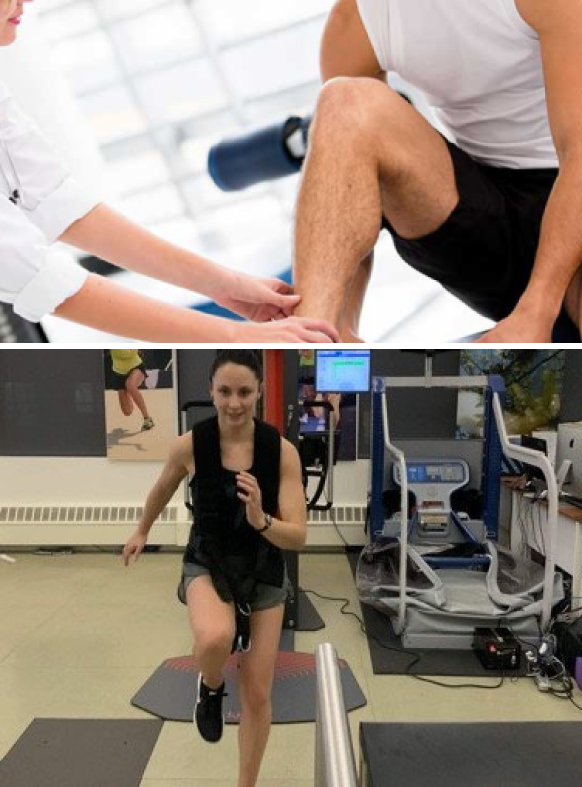
ForceFrame is a comprehensive system for accurately testing and training isometric strength in various muscle groups throughout the body. Most people – especially physically active people – develop muscle imbalances and compensation patterns over time that reduce movement efficiency.
ForceFrame lets us test individual muscle groups on both sides of the body for strength and symmetry. By testing individual muscle groups isometrically, we eliminate the potential for compensation of weak muscles that could be masked during compound exercise testing.
The results of each test appear as objective data on a screen in real time, helping us to identify and correct muscle imbalances, asymmetrical muscle strength, inefficient muscle firing patterns, and compensation patterns developed from past injuries. We then use that data to create personalized rehab programs, to boost performance and reduce injury risk.
The structures of the posterior kinetic chain are not isolated from one another – they work interdependently to provide static and dynamic stability. For example, proper function of the sacroiliac (SI) joints depends on the lumbodorsal fascia, which is connected to the pelvic ligamentous system. At the same time, the SI ligaments connect directly to the sacrotuberous ligaments, which attach directly to the proximal hamstring insertion. In other words, when one structure is injured, it can impact several others.
Lumbar problems generated by a herniated disc, piriformis syndrome or sciatica can impact the hamstrings, since those conditions all involve irritation or compression of the sciatic nerve. Even when issues affecting the lumbar discs are asymptomatic, they can cause over-activation of the hamstring muscles, or inhibit activation of the gluteal muscles, thereby overloading the hamstrings.
Simply treating hamstring pain without recognising the influence of other structures is negligent at best. Yet isolating body parts exemplifies modern medicine’s reductionist approach to hamstring pain. At NYDNRehab, our holistic integrative approach considers the complex interrelationships between various structures and systems. We don’t start treatment until we identify the exact cause of your hamstring pain.
At NYDNRehab, we treat the whole patient, not just your symptoms. We never use one-size-fits-all rehab protocols or antiquated recovery timelines. We believe that every injury is unique, and treatment should be based on a holistic approach that factors in the patient’s unique profile.
Once we have successfully pre-treated damaged tissues, we can begin one-on-one physical therapy to restore strength and stability, optimize mobility, and re-establish optimal neuromuscular pathways and muscle coordination patterns.
Your back-to-sports physical therapy protocol may include sport-specific training to optimize motor skills and restore peak athletic performance. We carefully monitor patient progress with ultrasound imaging to confirm complete recovery.
Advancements in technology are changing the game in rehabilitative medicine, enabling us to accelerate healing and restore performance at an unprecedented pace. The clinic at NYDNRehab features some of the most advanced therapeutic equipment currently available, and rarely found in private clinics.
Your hamstring physical therapy may incorporate advanced technologies:

Training and lifestyle factors both play a role in injury prevention. While hamstring injuries are common in sports, there are many things you can do to reduce your risk.

Hamstring injuries can take a long time to heal, and a wrong diagnosis can lead to the wrong treatment, drawing out the healing process. Once we arrive at a precise diagnosis, we select the most advanced rehabilitative technologies and therapies for your personalized treatment plan.
Technologies at NYDNRehab empower us to quantify your performance and monitor your progress, taking the guesswork out of injury rehab. We analyze your movement patterns from head to toe, to identify and eliminate any motor deficits. Our goal is to speed up healing and recovery so you can get back to your active lifestyle in the best shape ever.


Below is a prime example of how ultrasound can take the guesswork out of diagnosis.

A bad physical therapy experience is one of the primary causes of unnecessary surgery
In this instance, an athlete was originally diagnosed with minor quadriceps muscle strain and was treated for four weeks, with unsatisfactory results. When he came to our clinic, the muscle was not healing, and the patients’ muscle tissue had already begun to atrophy.
Upon examination using MSUS, we discovered that he had a full muscle thickness tear that had been overlooked by his previous provider. To mitigate damage and promote healing, surgery should have been performed immediately after the injury occurred. Because of misdiagnosis and inappropriate treatment, the patient now has permanent damage that cannot be corrected.
The most important advantage of Ultrasound over MRI imaging is its ability to zero in on the symptomatic region and obtain imaging, with active participation and feedback from the patient. Using dynamic MSUS, we can see what happens when patients contract their muscles, something that cannot be done with MRI. From a diagnostic perspective, this interaction is invaluable.
Dynamic ultrasonography examination demonstrating
the full thickness tear and already occurring muscle atrophy
due to misdiagnosis and not referring the patient
to proper diagnostic workup
Demonstration of how very small muscle defect is made and revealed
to be a complete tear with muscle contraction
under diagnostic sonography (not possible with MRI)

Complete tear of rectus femoris
with large hematoma (blood)

Separation of muscle ends due to tear elicited
on dynamic sonography examination


