HomeBlogHow To Approach Your Ankle Sprain Appropriately
How To Approach Your Ankle Sprain Appropriately
Inward Rolling vs Outward Rolling Sprains
All athletes who play sports with a heavy emphasis on running or pivoting are relatively likely to get an ankle sprain at some point in time. Generally, the most common type of ankle sprain will be one in which the ankle rolls inward. There might be a sharp pain on the outer tendons from an inner roll, but there usually isn’t much need for serious concern.
The sprain that requires the highest sense of urgency is when the foot rolls outward instead of an inward. An outward roll is usually serious enough to immediately incapacitate an athlete with distracting, immobilizing pain. It’s rarely severe enough to compromise an athlete’s entire career, but prompt ankle sprain treatment from a physician and proper rehabilitation techniques are essential for making a full recovery.
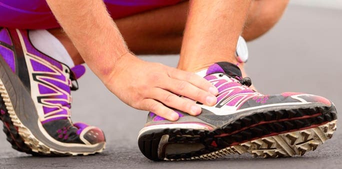
Ankle Sprain Swelling
Immediately following the occurrence of an outward rolling sprain, there will almost always be noticeable swelling. The swelling in your ankle after a sprain is caused by displaced ligaments. Even though human ankle ligaments are highly durable under normal conditions, hyper-extending or twisting them can cause ligaments to stretch out of place or completely snap.
Depending on the severity of the ligament damage, an ankle sprain can be categorized into one of three categories:
Grade 1: Hyper-extension of the ligaments without tearing
Grade 2: Some tearing, but not completely torn apart
Grade 3: The ligament has been completely torn
Elevation, Ice and Compression
The first recovery measure to take immediately after a sprain is to relieve it of unnecessary pressure. The athlete should take care to avoid putting their full body weight on the ankle for as long as possible, and if the pain is strong enough, using crutches may be a wise decision.
The athlete might benefit from icing the ankle periodically, which will take the swelling down considerably if done consistently enough. After the swelling has been slightly mitigated, an elastic bandage can be used to maintain the gradual swelling reduction.
Therapeutic Isometric Exercises
Once the pain has dwindled to a manageable level, the athlete may begin physical therapy by performing light isometric exercises. One isometric exercise can be performed by sitting in a chair (or the floor), straightening the leg outwards, and flexing ankle up and down. The athlete should avoid making any lateral ankle movements. Isometric exercises are designed to slowly facilitate a better rate of blood flow to the ankle.
Elastic band exercises
Another form of physical therapy can be performed by using elastic bands. Simply wrap the elastic band around the top of the foot, hold both ends in your hands, and slowly extend your leg out against the band’s resistance as far as possible. You may also tie the band’s ends around a heavy idle object and flex your ankle towards yourself, against the band’s resistance. Ten repetitions of both forms of elastic exercises should be more than enough.
In this instance, an athlete was originally diagnosed with minor quadriceps muscle strain and was treated for four weeks, with unsatisfactory results. When he came to our clinic, the muscle was not healing, and the patients’ muscle tissue had already begun to atrophy.
Upon examination using MSUS, we discovered that he had a full muscle thickness tear that had been overlooked by his previous provider. To mitigate damage and promote healing, surgery should have been performed immediately after the injury occurred. Because of misdiagnosis and inappropriate treatment, the patient now has permanent damage that cannot be corrected.
The most important advantage of Ultrasound over MRI imaging is its ability to zero in on the symptomatic region and obtain imaging, with active participation and feedback from the patient. Using dynamic MSUS, we can see what happens when patients contract their muscles, something that cannot be done with MRI. From a diagnostic perspective, this interaction is invaluable.
Dynamic ultrasonography examination demonstrating the full thickness tear and already occurring muscle atrophy due to misdiagnosis and not referring the patient to proper diagnostic workup
Demonstration of how very small muscle defect is made and revealed to be a complete tear with muscle contraction under diagnostic sonography (not possible with MRI)
Complete tear of rectus femoris with large hematoma (blood)
Separation of muscle ends due to tear elicited on dynamic sonography examination