Having Achilles tendonitis is definitely a setback, but it’s not the end of the world. A few weeks’ worth of rest and physical therapy will get you right back in the game.
The more you know about AT and how it occurs, the more likely you’ll be to take recovery seriously and prevent future injuries. Keep reading to find out what you can expect and to learn how you can accelerate the healing process.
AT Defined
It’s easier to understand AT if you know that the suffix “-itis” means inflammation.
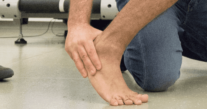
The Achilles tendon is a sturdy band of tissue that connects your heel to your calf muscles. Repeatedly pushing off from the ball of your foot puts undue stress on the tendon. Pain, tenderness, swelling, redness and heat that persist for several days are usually indications of AT.
The condition is quite common among athletes. Runners are especially susceptible; about half of all runners can expect to develop AT at some point in their active years.
Here are some possible causes of AT:
Over-exercising
Failing to give the tendon enough rest between workouts
Failing to properly stretch and warm up before activities
Suddenly increasing the duration or intensity of a training routine or workout
Be Good to Your Achilles Tendon
If you have AT, this is no time to be a hero. Playing through the pain will only exacerbate your injury and lead to other complications. AT never miraculously heals on its own. The sooner you make a firm commitment to doing everything that your doctor tells you to do, the faster you’ll recover and get back into action.
The first thing that you should do is suspend your preferred activity and rest the tendon. Depending on the severity of the problem, most experts recommend taking one to two months off. To stay in shape while you rehabilitate, you can take up low-impact exercises like water aerobics and cycling.
The next step is to start physical therapy. Here are some common methods that you can expect during treatment:
Hands-on treatment Your physical therapist will manipulate the tendon and ankle with his hands. The fibers in your tendon first start to heal with scar tissue, and manual therapy breaks up this weak tissue to improve motion and strength.
Focused exercise When your pain starts to subside, you’ll begin a regimen of therapeutic exercises. They’re designed to get the injured tissue back to optimal quality.
Pain control Ice and heat applications will help with pain. Your therapist may also incorporate electrical stimulation or ultrasound. Not only do these methods alleviate pain, but they also accelerate healing by increasing blood flow to the injured area.
Approach the healing process as though it were an important game or track meet. Play to win.
In this instance, an athlete was originally diagnosed with minor quadriceps muscle strain and was treated for four weeks, with unsatisfactory results. When he came to our clinic, the muscle was not healing, and the patients’ muscle tissue had already begun to atrophy.
Upon examination using MSUS, we discovered that he had a full muscle thickness tear that had been overlooked by his previous provider. To mitigate damage and promote healing, surgery should have been performed immediately after the injury occurred. Because of misdiagnosis and inappropriate treatment, the patient now has permanent damage that cannot be corrected.
The most important advantage of Ultrasound over MRI imaging is its ability to zero in on the symptomatic region and obtain imaging, with active participation and feedback from the patient. Using dynamic MSUS, we can see what happens when patients contract their muscles, something that cannot be done with MRI. From a diagnostic perspective, this interaction is invaluable.
Dynamic ultrasonography examination demonstrating the full thickness tear and already occurring muscle atrophy due to misdiagnosis and not referring the patient to proper diagnostic workup
Demonstration of how very small muscle defect is made and revealed to be a complete tear with muscle contraction under diagnostic sonography (not possible with MRI)
Complete tear of rectus femoris with large hematoma (blood)
Separation of muscle ends due to tear elicited on dynamic sonography examination