HomeBlogIliotibial Band (ITB) Syndrome: What To Know and What To Do About It?
Iliotibial Band (ITB) Syndrome: What To Know and What To Do About It?
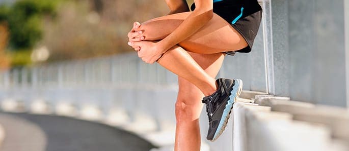
What Is Iliotibial Band Syndrome?
Iliotibial band syndrome or ITB syndrome is a family of symptoms that arise when the iliotibial band in the body is irritated or inflamed. The inflammation often happens in the impingement zone, when the knee is bent between 20 and 30 degrees.
Why Does ITB Syndrome Occur?
ITB syndrome occurs because the iliotibial band is constantly rubbed against the outside of the femur, or thigh bone that is close to the knee. This leads to inflammation, including inflammation of the bursa beneath the iliotibial band. The bursa is a sac filled with fluid found around most of the joints in the body. It reduces friction when the joints move.
ITB causes include the knee being repeatedly bent, which happens in sports such as rowing, running, bicycling. Overtraining, a sudden change in an athlete’s training regimen, the wrong footwear and the wrong running technique are also significant ITB causes.
Location Of ITB
The iliotibial band is a long, thick band of tissue that runs on the outside of a long bone in the leg called the femur. The iliotibial band runs from the pelvis to just below the knee. This is where it connects to the tibia and the fibula, which are the other long bones in the leg. The ITB also merges with the tensor fascia latae and gluteus maximus muscles.
What Does the ITB Do?
The ITB helps to straighten the knee, move the hip sideways and keep the legs stable when a person runs.
Top Six ITB Causes
Of the various ITB causes, sports doctors and physiotherapists have compiled a list of the top six that put people at higher risk for ITB. They are:
1. Motility Issues
Athletes, especially runners, who cannot move properly can put a great deal of pressure on the ITB. These runners do not take their leg backwards properly and rotate or turn the leg or foot out when their heel hits the ground. This puts them at higher risk for the syndrome.
2. Poor Technique
An athlete who jumps or lands with their foot turned out also puts a great deal of strain on the ITB. This particular motion by an athlete pulls their knee out and can stress the ITB where it connects to the knee. This is a particular issue for long-distance runners and marathoners.
3. The Athlete’s Own Biomechanics
Some athletes have what’s called patella mal-tracking, which means that the kneecap turns inward more than it should. This can put great stress on the iliotibial band. Not only this, but mal-tracking can lead to the kneecap becoming dislocated, which may, in turn, require surgery. People with patella mal-tracking can benefit from specific exercises to strengthen that area of the body.
4. Running Downhill
This type of running causes the knee to linger in the impingement zone, which increases the risk of ITB syndrome.
5. Tight Muscles
Tightness in the muscles can also increase the risk for the disorder, especially if it comes with shortening of the gluteus maximus muscles or the tensor fascia latae muscles. The athlete might also have overdeveloped muscles around their knee, which makes it more likely that ITB will occur at some point.
6. Weakened Muscles
People who have weak external rotator or abductor muscles around the hips or weak knee muscles are also more at risk for ITB. The risk is increased if the muscles are not only weak but the person is also fatigued when they attempt an athletic activity that involves working their knee or hip.
Symptoms of ITB
Initial signs of the syndrome may be a pain on the outside of the knee. The pain is especially severe when a person is walking down a flight of stairs. Running also worsens the pain, especially when the person is moving uphill. Some people describe the pain as a type of “hot pinching.”
Other symptoms include thickening or swelling of the tissue on the outside of the upper leg, and the knee also hurts when it’s bent or straightened. It is also more difficult to move the hips sideways. Some people hear snapping or popping around their knee area.
Diagnosing ITB Syndrome
The person’s doctor diagnoses the condition through taking note of the symptoms. They may also order an MRI or an ultrasound. These diagnostic tools also rule out other causes of knee pain such as injuries to the collateral ligament, anterior and posterior ligaments or meniscus tears.
ITB Treatment
IT band syndrome can almost always be rectified without the use of surgery. For most people, all that’s needed for the problem to be cleared up is rest and some pain medications. Afterwards, exercises and training should keep the syndrome at bay.
If a person believes they’re developing the symptoms of ITB, they should stop the activity that’s causing pain, employ the RICE procedure and call their doctor. The RICE procedure says to:
Rest the injured part
Ice the injured part. Do not apply ice directly to the area, but use an ice bag or even a bag of frozen peas. A cold compress can also be wrapped around the area for about 20 to 30 minutes. This will reduce the swelling, though some physical therapists actually claim that icing retards the healing process.
Compress the injured part.
Elevate the injured part.
If the symptoms haven’t gone away after a month or so of conservative ITB treatment, the doctor may give the patient corticosteroid injections and recommend massage, either with a physical therapist or by the patient him or herself. The doctor may also recommend such treatments as electrotherapy, which delivers a tiny electrical current to the area to stimulate healing. Ultrasound, phonophoresis and iontophoresis are also useful in alleviating the pain of ITB. Exercises are also an important part of ITB treatment. These include:
Stretching, Along With Icing
This exercise is controversial among some sports doctors and physical therapists because the iliotibial band is actually not supposed to stretch. Indeed, when it is stretched even a little bit it tears. Because of this, physiotherapists recommend that the glutes and the tensor fascia latae be stretched instead in support of the iliotibial band.
Massage
Self-massage up toward the hip and down toward the knee and even below the knee is also beneficial. The target is the trigger points, or painful points, in the gluteals and other muscles that involve the iliotibial band. Massage can also be done with equipment such as lacrosse balls, foam rollers or balls used in yoga tune-ups.
Strengthening the Muscles
People with weak muscles that can add to the risk of contracting ITB can benefit from strength training exercises. Physical therapists recommend beginning with open chain exercises. These include standing side-leg lifts. After a while, the patient can graduate to closed chain exercises such as single-leg step-downs and exercises to control the pelvic muscles. These exercises need to be done with both legs, and not just the one that’s injured. One of the exercises is the clam, which activates the hip flexors and glutes while supporting the stability in the abdominal and pelvic muscles. With a clam:
The person lies on their right slide while bending slightly at the hip and the knees. They extend their right arm so that it’s in line with the body, and lay their head on it. Then, they bend their left arm at the elbow and rest the left hand on the floor in front of them. The patient needs to keep their neck straight throughout the exercise.
Then, with the hips and shoulders lining up and their feet touching, they use their core to help lift the knee of the left leg while they rotate it at the hip.
Then, they lift the left knee as far as tit will go, while being careful to keep the hips aligned. Then, the patient slowly lowers their knee back to the start position, and repeats for the number of repetitions recommended by their physical therapist. They repeat the process with their right leg.
Other strengthening exercises are hip-hikes, sideways crab walks, step-downs and a hip control exercise using the wall as support.
Using a Compression Band
A compression band placed just above the affected knee helps to bring back circulation, which helps the body heal. After the compression band is applied, the patient holds on to something sturdy and performs deep squats. After 10 to 15 reps, the band is taken off.
Taping
Physical therapists claim that rock tape is excellent for people with ITB. Rock tape is a type of tape that interferes with the pain signals sent to the brain. When it’s applied, it causes the skin to wrinkle up, which takes pressure off of the tissue just beneath the skin. This improves the neurological, mechanical and fluid workings of the area.
When To Return to Sports
The decision on when to return to sports after ITB treatment varies from patient to patient. Some people are recovered after 6 weeks, but physical therapists warn that the patient should be able to properly perform closed and open chain exercises without pain before they return to running. Other therapists also say that faster running is better for an athlete who has just suffered a bout of ITB, because the knee spends less time in the impingement zone.
In this instance, an athlete was originally diagnosed with minor quadriceps muscle strain and was treated for four weeks, with unsatisfactory results. When he came to our clinic, the muscle was not healing, and the patients’ muscle tissue had already begun to atrophy.
Upon examination using MSUS, we discovered that he had a full muscle thickness tear that had been overlooked by his previous provider. To mitigate damage and promote healing, surgery should have been performed immediately after the injury occurred. Because of misdiagnosis and inappropriate treatment, the patient now has permanent damage that cannot be corrected.
The most important advantage of Ultrasound over MRI imaging is its ability to zero in on the symptomatic region and obtain imaging, with active participation and feedback from the patient. Using dynamic MSUS, we can see what happens when patients contract their muscles, something that cannot be done with MRI. From a diagnostic perspective, this interaction is invaluable.
Dynamic ultrasonography examination demonstrating the full thickness tear and already occurring muscle atrophy due to misdiagnosis and not referring the patient to proper diagnostic workup
Demonstration of how very small muscle defect is made and revealed to be a complete tear with muscle contraction under diagnostic sonography (not possible with MRI)
Complete tear of rectus femoris with large hematoma (blood)
Separation of muscle ends due to tear elicited on dynamic sonography examination