The foot and ankle is a complicated part of the body that has many moving parts that must work together to let us run, jump, stand on our toes, and walk. If one part of this group of muscles and bones gets injured, it often has a domino effect on the rest of the foot.
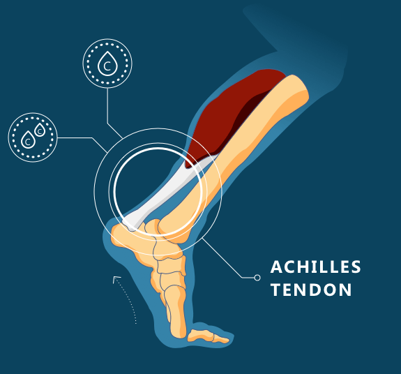
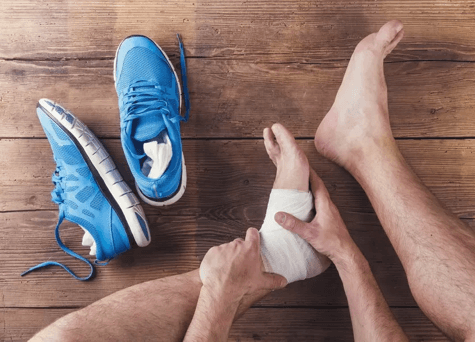
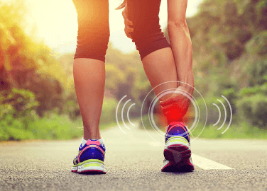
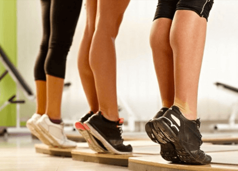
Attaching the calf muscle to the heel bone is a rubber band-like tendon called the Achilles. This tendon is used to move the foot forwards and backwards to help us have the mobility to jump, run, walk, and stand on the toes. Continuous physical activity and overuse can create painful inflammation of this area of the foot, known as tendinitis.
Almost the entire tendon consists of type-1 and type-3 collagen. Type-1 collagen, the stronger of the two, gives this tendon its strength. Unlike other tendons in the body, the Achilles rotates 90 degrees from its attached muscles to the back of the heel. This improves the efficiency in running, absorbing the shock of the heel hitting the ground, and letting the foot push off to take another step.
The Achilles tendon is a common place for an injury due to overuse. While it is not a weak tendon, there is a tremendous amount of strain put on it during physical activity. One study showed that 15% of military cadets suffered from an injury of their Achilles tendon after completing a six-week basic-training program. An Achilles tendon injury may be more common with age, because it weakens significantly as it gets older.

While injuries can be treated at home with stretches and exercises, it may be important to seek medical attention. If tendinitis continues to get worse, the tendon can tear, requiring medication and surgery.
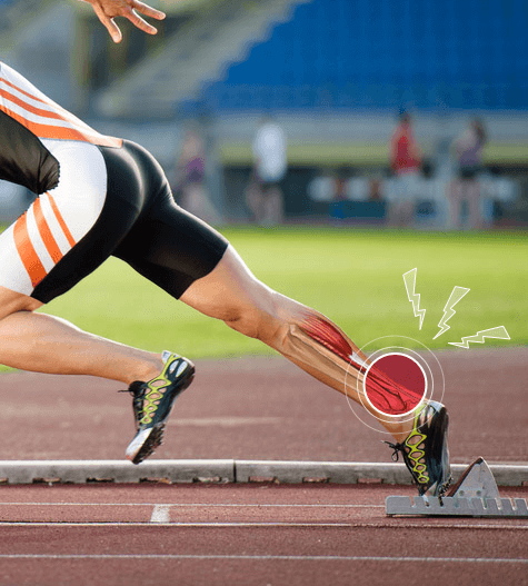
The Achilles tendon is strained and stretched when we push off to accelerate into running. The force put on this tendon can be up to seven times the weight of the average body, nearing the maximum amount of strain on Achilles tendon before rupturing.
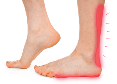
It is easy to feel this tendon. It is located above the heel on the back of the ankle and feels like a rubbery band of tissue. It is enveloped in a blanket of blood vessels which helps to keep it nourished.
Research actually confirms that the muscles attached to the Achilles tendon do very little work while pushing the body forward into acceleration. This leaves a lot of pressure and strain on the Achilles tendon to do all of the work and stretch while running and then return energy for the next step. The Achilles stores and returns energy to be able to continue to work throughout the running process. The more exercise that is done, the more the body learns to optimize this storage of energy to then be used to help the body accelerate.
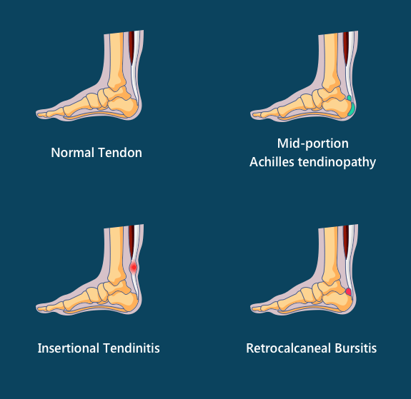
However, due to this intense strength that the Achilles stores, it also increased the chance of injury. There are different types of injuries that the Achilles can sustain. These fall into one of three categories, including insertional tendinitis, non-insertional tendinosis, and paratenonitis. Insertional tendinitis is the most common, so that is what will be discussed at length.
Insertional tendinitis is difficult to treat. It occurs when there is inflammation where the Achilles attaches to the heel. Here are some examples of what causes Achilles tendon injury, how to identify Achilles tendon injury, and how to recover Achilles tendon injury.
Insertional tendinitis most often occurs in people who have high arches and a tight Achilles tendon. A Haglund’s deformity is also a risk factor for this injury. This phenomenon is characterized by a bony mass on the back of the heel that causes the soft tissue around the tendon to become irritated when rubbing against the backs of shoes. This leads to retrocalcaneal bursitis. This is bursitis of the heel, which occurs when the fluid filled sacs that protect the bones and other moving parts of this area are aggravated.
While the most common causes of Achilles tendonitis are related to exercise and activity, other factors could also contribute to the risk of tendonitis. Infections that cause inflammation of the bursa or affect the white blood cells around the tendon, as well as rheumatoid arthritis are correlated with tendonitis.
Repetitive strain or use, however, is the most common cause. This includes exercising without warming up, straining the calf, making quick pivots or change of direction in sports, wearing poorly fitted shoes, and wearing high heeled shoes that stretch the bottom of the foot for a long period of time.
The development of this type of Achilles tendon injury once seemed obvious. It was thought that people with high arches and tight muscles put an increased strain on their Achilles tendon when the foot is bent upwards. While this seems logical, the opposite has proven to be true.
Researchers determined that the back of the Achilles tendon is less strained as the ankle is bent upwards, while the front of the tendon is exposed to less of stress. With insertional tendinitis, the back of the tendon is most often injured. This research shows the lack of stress on the front of the tendon may cause that portion of the tendon to weaken and eventually fail.
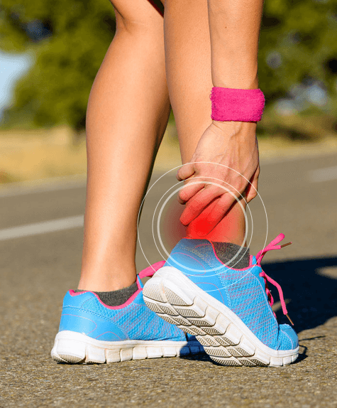
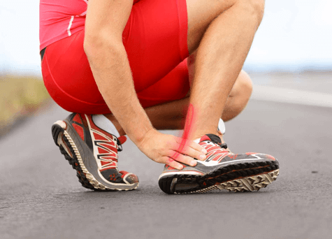
Achilles tendon injuries can come along with large amounts of pain. This pain is experienced all around the area of the foot, including along the back of the foot, the ankle, as well as above the heel. This is especially true when stretching the ankle or lifting up to stand on the balls of your feet. This pain may begin as being mild, yet gradually worsen.
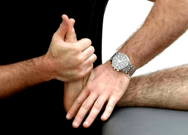
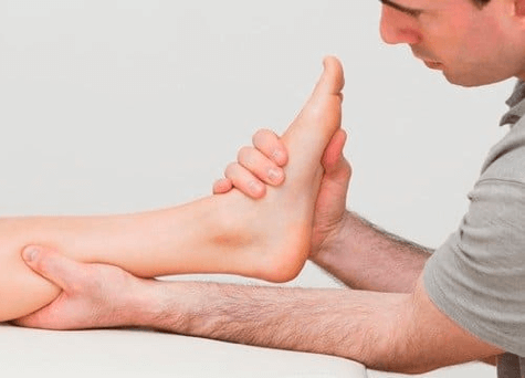
With an injury, it is difficult to engage the muscles around the tendon, especially if it is completely torn. In order to have this injury diagnosed, a doctor must give a thorough exam, watching the patient walk or jog while looking for issues that may have contributed to this injury.
If the tendon is ruptured, pain can come on quickly and be very severe. Some signs of rupturing include tenderness, stiffness, and swelling. You may even be able to hear the injury by the snapping or popping noise that it makes.
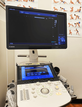
Aplio i800 ultrasound
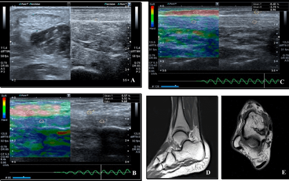
Insertional Achilles Tendonitis Sonoelastography
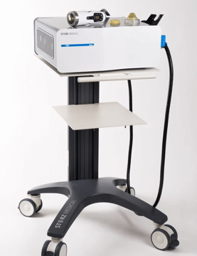
STORZ MEDICAL shockwave
It is important to strengthen the front tendons when treating insertional tendinitis. This can be done by performing specific exercises. Exercises are thought to create a slight pulsation that stimulates the production of collagen in the area in order to repair the tendon. Slowly raising and lowering yourself off your heels creates a slight muscle pulsation that places a greater amount of strain on the front portion of the tendon, causing it to strengthen.
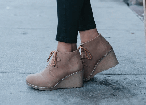
When these injuries happen in people with high arches, the best treatment is the use of slighted wedged shoes. Having heel lifts increases the activity in the muscles surrounding the Achilles tendon, which helps to keep them strong. Anyone who is at a high risk for an Achilles tendon injury or is strongly trying to prevent it can use these special shoes to keep the muscles in the foot strong. A night brace should also be considered for those who experience pain in the morning.
Lengthening the muscles in the heel with gentle stretches along with a deep tissue massage can relieve some pain. Mild stretching is sufficient, as too much stretching may cause a re-injury. All healing of an Achilles tendon injury should be performed gently.
Prior to stretching, massage the calf muscles and the back of the ankle in order to help lengthen and relax the muscles as fast as possible. Deep-tissue massage increases the range of motion of the muscles more effectively.
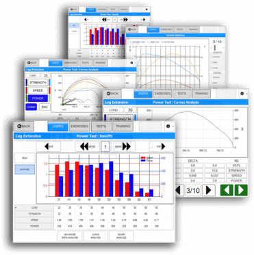
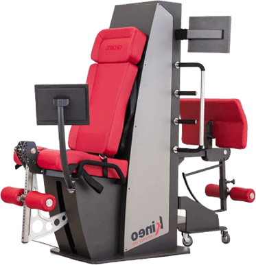
Tendons heal by loading them. The Kineo intelligent load system was developed by professional soccer teams for treatment and prevention of various tendon disorders and ACL tears.
At our clinic, we have extensive experience in Achilles Tendon Injury relief.


Research shows that by far the greatest results were achieved with any type of Achilles tendinopathy by combining ESWT with eccentric loading exercises
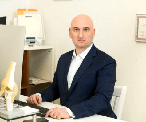
Dr.Kalika has revolutionized Achilles tendon care by using high resolution diagnostic ultrasonography for structural diagnosis, combined with gait and motion analysis technology. The NYDNRehab motion and gait analysis lab is the only private lab in the US to feature research-grade technologies found only in the world’s top research labs, and made available to patients in our private clinic. Dr.Kalika’s modern approach to athletic Achilles tendon injuries has put him on the radar of some of the world’s top distance runners, pro athletes and professional ballet dancers.
 Dr. Yuri Brosgol
MD
Dr. Yuri Brosgol
MD
 Dr. Michael Goynatsky
DPT
Dr. Michael Goynatsky
DPT
 Dr. Daniela Escudero
DPT
Dr. Daniela Escudero
DPT
 Dr. Michelle Agyakwah
DC
Dr. Michelle Agyakwah
DC
 Dr. Tatyana Kapustina
L. Ac.
Dr. Tatyana Kapustina
L. Ac.

Verified Expert Profiles
Dr. Lev Kalika is a world-recognized expert in musculoskeletal medicine. with 20+ years of clinical experience in diagnostic musculoskeletal ultrasonography, rehabilitative sports medicine and conservative orthopedics. In addition to operating his clinical practice in Manhattan, he regularly publishes peer-reviewed research on ultrasound-guided therapies and procedures. He serves as a peer reviewer for Springer Nature.
Dr. Kalika is an esteemed member of multiple professional organizations, including:
Below is a prime example of how ultrasound can take the guesswork out of diagnosis.

A bad physical therapy experience is one of the primary causes of unnecessary surgery
In this instance, an athlete was originally diagnosed with minor quadriceps muscle strain and was treated for four weeks, with unsatisfactory results. When he came to our clinic, the muscle was not healing, and the patients’ muscle tissue had already begun to atrophy.
Upon examination using MSUS, we discovered that he had a full muscle thickness tear that had been overlooked by his previous provider. To mitigate damage and promote healing, surgery should have been performed immediately after the injury occurred. Because of misdiagnosis and inappropriate treatment, the patient now has permanent damage that cannot be corrected.
The most important advantage of Ultrasound over MRI imaging is its ability to zero in on the symptomatic region and obtain imaging, with active participation and feedback from the patient. Using dynamic MSUS, we can see what happens when patients contract their muscles, something that cannot be done with MRI. From a diagnostic perspective, this interaction is invaluable.
Dynamic ultrasonography examination demonstrating
the full thickness tear and already occurring muscle atrophy
due to misdiagnosis and not referring the patient
to proper diagnostic workup
Demonstration of how very small muscle defect is made and revealed
to be a complete tear with muscle contraction
under diagnostic sonography (not possible with MRI)

Complete tear of rectus femoris
with large hematoma (blood)

Separation of muscle ends due to tear elicited
on dynamic sonography examination


