
Despite technology’s many advantages for enhancing human life, the ubiquitous use of computers and mobile devices is taking a toll on our bodies. Too much time spent sitting and slouching over keyboards and mobile devices, and too little physical activity in general, cause strain and imbalances that lead to pain and injury.
Functional movement allows you to perform everyday tasks efficiently, without pain or discomfort. Integrative motion analysis helps us pinpoint underlying issues that restrict functional movement and inhibit mobility. We are then able to create corrective treatment programs that restore fluid functional movement and improve quality of life.
Despite our similarities, human beings have unique movement patterns and distinct anatomical characteristics. Treatment is often generalized, with exercise prescriptions based on what works in a research setting. IMA lets us look at the unique muscle activation patterns of each individual patient, so we can provide the most effective personalized treatment.
Integrative motion analysis (IMA) provides tools for quantifying various movement parameters, to assess dysfunction and track recovery. In human movement, many factors come into play that define our quality of movement:
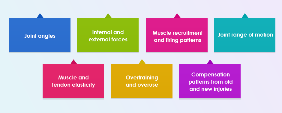
Most of those factors cannot be assessed with the naked eye, and require advanced technological tools to analyze and quantify. A thorough assessment with IMA is the best way to assess, quantify and restore functional movement.

IMA begins with a biomechanical examination to help us determine if a body part is functioning in an abnormal way. The exam has 3 goals:
The biomechanical exam includes assessments for function and joint range of motion, and muscle testing for tension and strength. Once we have all the information we need, we can craft a personalized treatment plan for our patient, to restore normal function and enhance functional movement.
IMA uses multiple tools to get a comprehensive picture of how well your body moves, and to identify kinematic issues that keep you from moving with optimal efficiency.

Tools for IMA include:
The sophisticated technology we use at NYDNRehab is most often used in academia for research. It is rarely found in a private clinic, or available to private patients. We have spent years studying, innovating and integrating our equipment, to improve the functional performance of patients and athletes. Our technological tools are paired with custom software that collects and analyzes patient data, providing a quantified baseline to help us measure patient progress.
Injuries from sports and overuse often arise from faulty movement mechanics. After an injury, your neuromuscular system intuitively protects the damaged tissues by shifting force loads to other structures. In time, compensation patterns can become habitual, creating dysfunctional movement, long after the tissues have healed.
State-of-the-art motion analysis identifies compensation patterns and helps us develop an individualized treatment protocol. IMA eliminates time wasted on ineffective rehab, and speeds up the patient’s return to activity.
We use IMA to diagnose and retrain the following musculoskeletal injuries:
IMA gives us precise quantitative data, taking the guesswork out of rehab and individualizing the rehabilitation process.
In addition to assessing movement, IMA helps us retrain muscle activation patterns using real-time feedback. After an injury, neuroplastic changes to the brain and central nervous system can cause compensation patterns that interfere with coordinated muscle recruitment. It is not enough to simply retrain the muscles. We need to retrain the brain to access the right muscles at the right time.
All too often, rehab programs focus on healing damaged tissues and strengthening the surrounding muscles without addressing brain neuroplasticity. Failure to retrain the brain along with the muscles dramatically increases the risk of reinjury and undermines performance.
In addition to assessing movement, IMA helps us retrain muscle activation patterns using real-time feedback. After an injury, neuroplastic changes to the brain and central nervous system can cause compensation patterns that interfere with coordinated muscle recruitment. It is not enough to simply retrain the muscles. We need to retrain the brain to access the right muscles at the right time.
Resources:
Einarsson, Elinar. “Electromyography and Clinical Reasoning; Why do exercise treatments sometimes fail and how can we overcome this?” ASPETAR Sports Medicine Journal 5.1 (2016).

Verified Expert Profiles
Dr. Lev Kalika is a world-recognized expert in musculoskeletal medicine. with 20+ years of clinical experience in diagnostic musculoskeletal ultrasonography, rehabilitative sports medicine and conservative orthopedics. In addition to operating his clinical practice in Manhattan, he regularly publishes peer-reviewed research on ultrasound-guided therapies and procedures. He serves as a peer reviewer for Springer Nature.
Dr. Kalika is an esteemed member of multiple professional organizations, including:
Below is a prime example of how ultrasound can take the guesswork out of diagnosis.

A bad physical therapy experience is one of the primary causes of unnecessary surgery
In this instance, an athlete was originally diagnosed with minor quadriceps muscle strain and was treated for four weeks, with unsatisfactory results. When he came to our clinic, the muscle was not healing, and the patients’ muscle tissue had already begun to atrophy.
Upon examination using MSUS, we discovered that he had a full muscle thickness tear that had been overlooked by his previous provider. To mitigate damage and promote healing, surgery should have been performed immediately after the injury occurred. Because of misdiagnosis and inappropriate treatment, the patient now has permanent damage that cannot be corrected.
The most important advantage of Ultrasound over MRI imaging is its ability to zero in on the symptomatic region and obtain imaging, with active participation and feedback from the patient. Using dynamic MSUS, we can see what happens when patients contract their muscles, something that cannot be done with MRI. From a diagnostic perspective, this interaction is invaluable.
Dynamic ultrasonography examination demonstrating
the full thickness tear and already occurring muscle atrophy
due to misdiagnosis and not referring the patient
to proper diagnostic workup
Demonstration of how very small muscle defect is made and revealed
to be a complete tear with muscle contraction
under diagnostic sonography (not possible with MRI)

Complete tear of rectus femoris
with large hematoma (blood)

Separation of muscle ends due to tear elicited
on dynamic sonography examination


