
Physically active people can have a plethora of injuries over the course of a lifetime, and as much as they hurt at the time, many injuries are forgotten, once the pain subsides and function returns. ACL injuries and tears are especially common for young athletes, dancers and fitness lovers, and even with reconstructive surgery and rehab, they can come back to haunt you, years down the road.
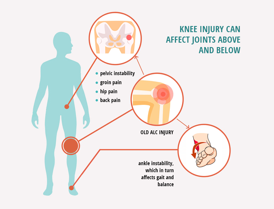
Because of the knee joint’s location and function in the body’s lower kinetic chain, a knee injury can affect joints above and below. A knee that is not fully rehabbed after injury, or that deteriorates over time, can alter the way body weight is distributed over the ankle and foot, causing misalignment that leads to ankle instability, which in turn affects gait and balance, and increases injury risk during physical activity.
In the same way, a dysfunctional knee can translate upward, affecting the hip and misaligning the spine. This can lead to hip pain and instability, groin pain, pelvic instability and back pain. While many people write these issues off as the consequences of aging, they are in reality a consequence of injury.
The good news is that physical therapy can help improve knee function, even years after an injury, and dramatically reduce pain, dysfunction and instability throughout the body.
New research about the long-term consequences of knee injury is shedding light on the extent of the initial damage done to the tissues surrounding the knee, and how immediate treatment affects long-term outcomes.

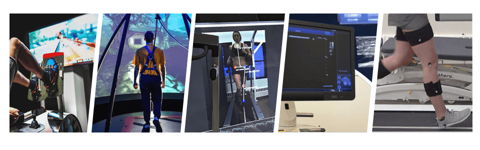
If you have an old ACL rupture or knee injury from younger, more active years, having a thorough gait and biomechanical analysis now could help prevent future problems.
At NYDNRehab, we go beyond treating the knee and its surrounding structures after an ACL injury. We take rehab to the next level by reestablishing the relationship between the muscles and the brain, to ensure fully restored function.
The team at NYDNRehab has rehabilitated over 100 patients with torn ACLs over the past six years. All had ACL tear treatment without surgery using our meticulous approach and advanced modern technology. Our results closely reflect those of various research studies. Only 20 of our patients were unable to return to sports, and subsequently went for surgical ACL repair.
At NYDNRehab, we go beyond treating symptoms of pain and dysfunction. Our advanced technologies and innovative treatment methods enable us to identify the underlying causes, collect baseline data, and develop rehabilitation protocols that restore neuromuscular and neurocognitive function. Our end goal is to eliminate pain and dysfunction, so our patients can enjoy the very best quality of life.

Verified Expert Profiles
Dr. Lev Kalika is a world-recognized expert in musculoskeletal medicine. with 20+ years of clinical experience in diagnostic musculoskeletal ultrasonography, rehabilitative sports medicine and conservative orthopedics. In addition to operating his clinical practice in Manhattan, he regularly publishes peer-reviewed research on ultrasound-guided therapies and procedures. He serves as a peer reviewer for Springer Nature.
Dr. Kalika is an esteemed member of multiple professional organizations, including:
Below is a prime example of how ultrasound can take the guesswork out of diagnosis.

A bad physical therapy experience is one of the primary causes of unnecessary surgery
In this instance, an athlete was originally diagnosed with minor quadriceps muscle strain and was treated for four weeks, with unsatisfactory results. When he came to our clinic, the muscle was not healing, and the patients’ muscle tissue had already begun to atrophy.
Upon examination using MSUS, we discovered that he had a full muscle thickness tear that had been overlooked by his previous provider. To mitigate damage and promote healing, surgery should have been performed immediately after the injury occurred. Because of misdiagnosis and inappropriate treatment, the patient now has permanent damage that cannot be corrected.
The most important advantage of Ultrasound over MRI imaging is its ability to zero in on the symptomatic region and obtain imaging, with active participation and feedback from the patient. Using dynamic MSUS, we can see what happens when patients contract their muscles, something that cannot be done with MRI. From a diagnostic perspective, this interaction is invaluable.
Dynamic ultrasonography examination demonstrating
the full thickness tear and already occurring muscle atrophy
due to misdiagnosis and not referring the patient
to proper diagnostic workup
Demonstration of how very small muscle defect is made and revealed
to be a complete tear with muscle contraction
under diagnostic sonography (not possible with MRI)

Complete tear of rectus femoris
with large hematoma (blood)

Separation of muscle ends due to tear elicited
on dynamic sonography examination


