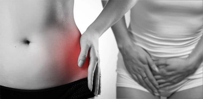
A recent study of professional soccer players found 20% of all their injuries involved the areas near the thigh or groin. This finding proved the theory that athletes in sports requiring quick footwork, such as soccer or lacrosse, tend to suffer from injury to the hips and groin. This injury often happens because of overuse. The pain associated with hip and groin injuries can be quite severe, so it is important to diagnose the problem to obtain the correct treatment. There are many possible causes of hip and groin pain.
One common cause of hip and groin pain is a strain or tear of one or several muscles. Strains often occur due to overuse. A strain often occurs in these following areas:
The last item on the list is called IT band syndrome, and this is caused by overuse of connective tissues. There is currently no exact way to diagnose this kind of injury. However, a strain can be detected through a trial-and-error method of asking the patient to contract each muscle while pushing against resistance. This method, along with imaging, is one of the best ways to determine the cause of pain.
Pain that is joint-related often occurs along the crease of the hip or in the groin line. The labrum refers to the circle of cartilage that surrounds the hip joint. A tear in this cartilage can lead the death of bone tissue. If an individual has been treated with steroidal medications, this condition should be considered when diagnosing the cause pain in the hip or groin. A combination of a thorough physical examination and x-rays can often effectively diagnose most causes of pain in the hip region, but in the case of a labral tear, a doctor may order an MRI arthrogram to study the affected area. Dye will be injected directly into this joint before imaging begins.
Whenever a patient complains of persistent discomfort in the groin or hip area, a healthcare professional could guess that there may be a stress fracture at the source of the pain. Stress fractures occur when muscles are too exhausted to absorb shock. If the patient reports worsening pain when he or she puts weight on one leg, there is even more of a chance that there is a stress fracture. This suspicion can be confirmed through a bone scan or MRI. The usual sites for this kind of fracture are the femur and the pubic bone. A fracture of one of these bones can be particularly serious depending on its exact location. A fracture at the part of femur that is near the hip joint requires vigorous treatment, as it can be debilitating. Often, abrupt changes in an athlete’s training can put him or her at risk for a stress fracture.
Bursitis is one of the most common cause of pain in the hip and groin. This condition occurs when a bursa becomes inflamed. Healthy bursae reduce friction between bones and tendons or between muscles, but repetitive motions can irritate bursae and contribute to bursitis. If an athlete notices pain on the outside of the hip area, this may be trochanteric bursitis. If the pain is located in the groin or in the thigh, this is known as psoas bursitis, which affects the muscle that connects the femur to the lumbar vertebrae. Gently pressing on each area and observing the patient’s pain response should be able to help find where the bursitis is located.
Nerve injury can be a cause of pain as well, and it can be one of the hardest injuries to diagnose. People with nerve-related pain may complain of tingling or a burning sensation in the hip or groin areas. If a patient has a history of cycling, this may mean that she or she could have injured nerves due to overusing them.
There are many other conditions that can cause pain in the hip or groin area. Hernias within or above the groin can cause discomfort, as can irritation of the front pubic joint. Advanced urinary tract infections and other gynecological infections can cause pain as well. The occurrence of uterine tissue outside of the uterus is known as endometriosis, and this can cause discomfort near the hip or groin. Inflammation of the testicles or prostate can cause discomfort, and any swelling can possibly be due to testicular cancer.
Are you looking for a high-end clinic to undergo physical therapy in NYC? Visit our main page to find out valuable information.

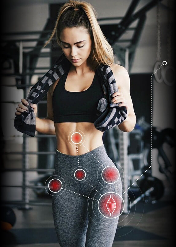
A clinical exam and diagnostic ultrasound imaging can help your therapist pinpoint the exact location and cause of your hip and groin pain.
Ultrasound enables you and your therapist to view the hip and groin region in real time, while in motion. In addition to ultrasound, video gait analysis can help us identify faulty movement mechanics that contribute to hip and groin pain. Once the exact cause is determined, an effective treatment plan can be initiated.
Please explore more advanced diagnostic option unavailable anywhere else:

Hip dysfunction and pain can be a complex issue due to interactions of the trunk, pelvis, low back, groin and hip joint. Physical therapy and rehabilitation that is based only on subjective clinical analysis often addresses the symptoms without resolving the underlying cause.
At NYDNRehab, our groundbreaking motion analysis technology and high resolution diagnostic ultrasonography have enabled us to develop a battery of tests that perfectly reveal the dynamic functional pathology of the hip joint and pelvis. Our tests are evidence-based protocols that are considered to be the gold standard in the world of research.
Our testing protocol includes:
Combined lumbopelvic hip stability test using DLEST methodology with C.A.R.E.N., our computer assisted rehab environment
Hip joint stability test using DLEST methodology with C.A.R.E.N.
3D star excursion banner test (SEBT) for assessing the involvement of the hip joint and muscles in postural stability
3D gait or running analysis
3D kinematic joint angle analysis during a squat, lunge, drop jump and pelvis on hip rotation
Rehabilitative ultrasonography for viewing intrinsic hip stabilizing muscle activation patterns

Verified Expert Profiles
Dr. Lev Kalika is a world-recognized expert in musculoskeletal medicine. with 20+ years of clinical experience in diagnostic musculoskeletal ultrasonography, rehabilitative sports medicine and conservative orthopedics. In addition to operating his clinical practice in Manhattan, he regularly publishes peer-reviewed research on ultrasound-guided therapies and procedures. He serves as a peer reviewer for Springer Nature.
Dr. Kalika is an esteemed member of multiple professional organizations, including:
Below is a prime example of how ultrasound can take the guesswork out of diagnosis.

A bad physical therapy experience is one of the primary causes of unnecessary surgery
In this instance, an athlete was originally diagnosed with minor quadriceps muscle strain and was treated for four weeks, with unsatisfactory results. When he came to our clinic, the muscle was not healing, and the patients’ muscle tissue had already begun to atrophy.
Upon examination using MSUS, we discovered that he had a full muscle thickness tear that had been overlooked by his previous provider. To mitigate damage and promote healing, surgery should have been performed immediately after the injury occurred. Because of misdiagnosis and inappropriate treatment, the patient now has permanent damage that cannot be corrected.
The most important advantage of Ultrasound over MRI imaging is its ability to zero in on the symptomatic region and obtain imaging, with active participation and feedback from the patient. Using dynamic MSUS, we can see what happens when patients contract their muscles, something that cannot be done with MRI. From a diagnostic perspective, this interaction is invaluable.
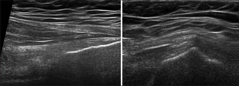
Dynamic ultrasonography examination demonstrating
the full thickness tear and already occurring muscle atrophy
due to misdiagnosis and not referring the patient
to proper diagnostic workup
Demonstration of how very small muscle defect is made and revealed
to be a complete tear with muscle contraction
under diagnostic sonography (not possible with MRI)

Complete tear of rectus femoris
with large hematoma (blood)

Separation of muscle ends due to tear elicited
on dynamic sonography examination


