August 14, 2023
 Ankle sprains are very common injuries, and people who suffer from them may suffer ongoing problems, including recurring sprains. Each year, seven ankle sprains occur for every 1,000 people. Up to address dysfunctions of the joints may result in better outcomes.
Joint dysfunctions that can occur with inversion ankle sprains, which happen when the outer ligaments around the ankle are over-stretched.
Researchers have studied the ankles of people who suffer from recurring ankle sprains in order to suffer from re-injuries. They have found that people may suffer from a variety of joint dysfunctions, including:
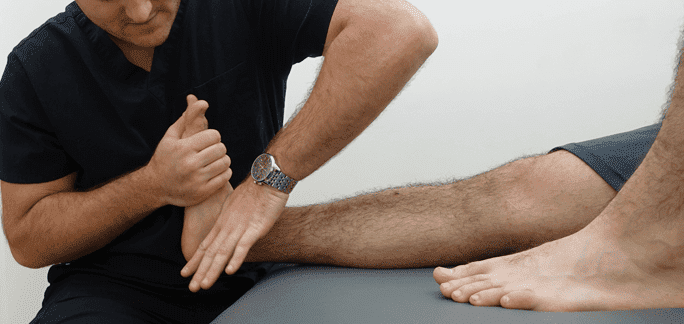
By directly addressing all existing joint dysfunctions, therapists who use manual therapy in the treatment of ankle sprains may be able to minimize the risk of ongoing ankle problems.
While several studies have demonstrated the potential short-term benefits of using manual manipulation for the treatment of sprained ankles, the long-term benefits were unknown until a 2013 study. In that study, seventy-four people were randomly placed in one of two groups, including one that performed home exercises as directed by their physical therapists and one in which the patients received manual therapy in addition to performing home exercises as directed by their therapists.
The patients’ ankles were given a baseline assessment prior to the study’s start. Their ankles were then reassessed at four weeks and at six months using multiple scales, including the Foot and Ankle Ability Measure ADL and sports subscales, the Lower Extremity Functional Scale, the NPRS and the 15-point global scale of change.
The participants in the home exercise group saw their physical therapists for 30 minutes once a week for four weeks. They were each given research-based exercise routines to manipulate their joints during their sessions. The people also completed the same home exercise regimens that the home exercise group participants did.
Sixty-five of the original 74 participants completed both their four-week and six-month follow-up appointments. The people who had received manual therapy in addition to the 15.6 percent recurrence rate among those who did not receive manual therapy.
By addressing joint dysfunctions with manual therapy, patients may enjoy better outcomes and fewer re-injuries of their ankles. People who have suffered inversion ankle sprains may want tocols.

Verified Expert Profiles
Dr. Lev Kalika is a world-recognized expert in musculoskeletal medicine. with 20+ years of clinical experience in diagnostic musculoskeletal ultrasonography, rehabilitative sports medicine and conservative orthopedics. In addition to operating his clinical practice in Manhattan, he regularly publishes peer-reviewed research on ultrasound-guided therapies and procedures. He serves as a peer reviewer for Springer Nature.
Dr. Kalika is an esteemed member of multiple professional organizations, including:
Below is a prime example of how ultrasound can take the guesswork out of diagnosis.

A bad physical therapy experience is one of the primary causes of unnecessary surgery
In this instance, an athlete was originally diagnosed with minor quadriceps muscle strain and was treated for four weeks, with unsatisfactory results. When he came to our clinic, the muscle was not healing, and the patients’ muscle tissue had already begun to atrophy.
Upon examination using MSUS, we discovered that he had a full muscle thickness tear that had been overlooked by his previous provider. To mitigate damage and promote healing, surgery should have been performed immediately after the injury occurred. Because of misdiagnosis and inappropriate treatment, the patient now has permanent damage that cannot be corrected.
The most important advantage of Ultrasound over MRI imaging is its ability to zero in on the symptomatic region and obtain imaging, with active participation and feedback from the patient. Using dynamic MSUS, we can see what happens when patients contract their muscles, something that cannot be done with MRI. From a diagnostic perspective, this interaction is invaluable.
Dynamic ultrasonography examination demonstrating
the full thickness tear and already occurring muscle atrophy
due to misdiagnosis and not referring the patient
to proper diagnostic workup
Demonstration of how very small muscle defect is made and revealed
to be a complete tear with muscle contraction
under diagnostic sonography (not possible with MRI)

Complete tear of rectus femoris
with large hematoma (blood)

Separation of muscle ends due to tear elicited
on dynamic sonography examination


