Diastasis recti is a common complaint among new mothers. When you think about it, it is truly amazing how the human female body is able to adapt to the needs of a growing fetus. As your baby grows and approaches full term, something’s gotta give, and it is often the linea alba, the tough fibrous band that connects the left and right sides of your abs. Pressure on the linea alba may cause it to split apart, leaving a gap in your abdominal wall.
The postpartum period is often called the “fourth trimester” because it is a time of adapting for both mom and baby. Your baby is learning to live outside the cozy environment of the womb, and your body is healing and recovering from pregnancy and childbirth. For many new mothers, that means restoring the integrity of an abdominal wall that has been stretched and split during pregnancy.
Sadly, many women are totally unaware of diastasis recti, and they are surprised when their body does not bounce back to its pre-pregnancy shape after childbirth. Most books and videos on pregnancy fail to mention it, and for that matter, women are given very little information about postpartum self-care at all.
When the two sides of your abs separate, you lose support for your back and internal organs. Just imagine buttoning the waistband of your jeans but leaving the zipper open — a little bit of tummy is going to bulge out.
Signs of diastasis recti include:
Being unaware of your diastasis recti both during and after pregnancy can actually make the condition worse, so it is important to identify and proactively treat it early on.
Many women panic when they first discover diastasis recti, and surgery often comes to mind. However, most cases of diastasis recti can be resolved without surgical intervention with the help of specialized physical therapy exercises. Resolving diastasis recti through exercise will make you stronger and help you prevent the condition in future pregnancies.
The linea alba is mostly made up of collagen, which your body produces to make repairs. When treated with specific therapeutic exercises, the linea alba can begin to mend and the gap eventually closes, restoring the integrity of your abdominal region.
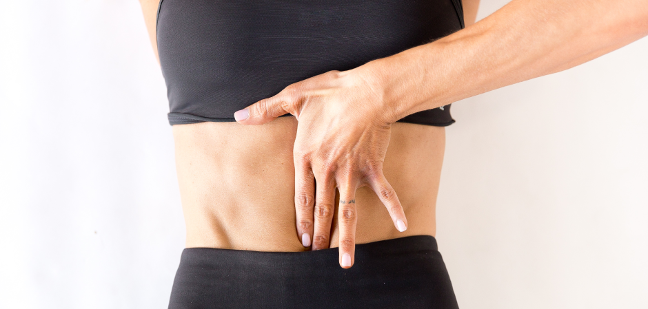
The gap caused by diastasis recti is called the inter-recti distance, or IRD. In order to assess the effectiveness of corrective exercises, the IRD should be measured as a baseline prior to treatment, and then periodically as treatment progresses. Normally, IRD is measured in three places:
While there is no consensus as to the exact distance for obtaining optimal measurements, some researchers suggest that the patient’s height and the length of the linea alba should be taken into consideration, and that the measurement site should be calculated as a percentage of length. Regardless of the distance above or below the belly button, it is important to measure at the exact same site each time, to assess the degree of healing.
Traditionally, clinicians measure diastasis recti with the patient in a crook-lying supine position, with the head elevated and knees bent. However, a recent study by Gillard et al (2018) argued that the gap is more accurately measured in weight-bearing postures while sitting or standing, where the lateral abdominal muscles are activated to provide stability, providing a better idea of the progress of functional recovery.
The research team further advocated for the use of ultrasound imaging as a superior way of measuring healing progress. Ultrasound enables the clinician to not only accurately measure gap distance, but to detect structural changes in the linea alba.
If you suspect you have diastasis recti, it is important to immediately seek help to resolve it. The women’s health specialists at NYDNR use diagnostic ultrasound to detect and measure diastasis recti. We apply our experience and knowledge to help you recover through physical therapy exercises, designed specifically for diastasis recti. Contact us today, and get your body on the road to recovery from pregnancy and childbirth with NYDNR.
Gillard, Samantha, et al. “Effects of posture and anatomical location on inter-recti distance measured using ultrasound imaging in parous women.” Musculoskeletal Science and Practice 34 (2018): 1-7.

Verified Expert Profiles
Dr. Lev Kalika is a world-recognized expert in musculoskeletal medicine. with 20+ years of clinical experience in diagnostic musculoskeletal ultrasonography, rehabilitative sports medicine and conservative orthopedics. In addition to operating his clinical practice in Manhattan, he regularly publishes peer-reviewed research on ultrasound-guided therapies and procedures. He serves as a peer reviewer for Springer Nature.
Dr. Kalika is an esteemed member of multiple professional organizations, including:
Below is a prime example of how ultrasound can take the guesswork out of diagnosis.

A bad physical therapy experience is one of the primary causes of unnecessary surgery
In this instance, an athlete was originally diagnosed with minor quadriceps muscle strain and was treated for four weeks, with unsatisfactory results. When he came to our clinic, the muscle was not healing, and the patients’ muscle tissue had already begun to atrophy.
Upon examination using MSUS, we discovered that he had a full muscle thickness tear that had been overlooked by his previous provider. To mitigate damage and promote healing, surgery should have been performed immediately after the injury occurred. Because of misdiagnosis and inappropriate treatment, the patient now has permanent damage that cannot be corrected.
The most important advantage of Ultrasound over MRI imaging is its ability to zero in on the symptomatic region and obtain imaging, with active participation and feedback from the patient. Using dynamic MSUS, we can see what happens when patients contract their muscles, something that cannot be done with MRI. From a diagnostic perspective, this interaction is invaluable.
Dynamic ultrasonography examination demonstrating
the full thickness tear and already occurring muscle atrophy
due to misdiagnosis and not referring the patient
to proper diagnostic workup
Demonstration of how very small muscle defect is made and revealed
to be a complete tear with muscle contraction
under diagnostic sonography (not possible with MRI)

Complete tear of rectus femoris
with large hematoma (blood)

Separation of muscle ends due to tear elicited
on dynamic sonography examination


