Patients often come to the doctor for treatment of a back ailment, and two of the most common are sciatica and herniated discs. When leg pain, numbness or weakness occurs, most clinicians and patients will turn to the same diagnostic tool: an MRI scan. The assumption is simple: if imaging can demonstrate the problem, it should also be able to reveal the optimum treatment strategy. But the connection between the MRI and patient’s symptoms is much more complex.
Lumbar disc herniations are the result of the soft inner portion of the disc in the spine bulging outwards and possibly damaging the surrounding nerve roots. This irritation may lead to radiating pain, numbness or weakness down one or both legs (sciatica, or more specifically lumbar radiculopathy). Although this diagnosis is common, determining the need for surgery or conservative treatment is a major clinical dilemma.

Introduction
MRI is a common test ordered for patients with sciatica, as it gives detailed pictures of disc herniation, nerve root compression and the anatomy of the spinal canal. However, new studies show that MRI data is not enough and can sometimes be misleading when deciding on treatment. In particular, MRI scans are often abnormal even in individuals with no symptoms at all. This article reviews a major clinical study and the overall evidence base to assist patients and clinicians in understanding the limitations and value of MRI in the diagnosis of sciatica.
Sciatica is a condition characterized by pain that travels down the length of the sciatic nerve from the lower back through the hips and buttocks down each leg. It is not a diagnosis, but a group of symptoms resulting from inflammation or compression of one or more nerve roots in the lumbar spine.
Symptoms of sciatica are:

Unlike lower back pain in general, the main complaint of people with sciatica is the pain in the leg, not the back. Symptoms may be mild and intermittent or severe and disabling and will differ widely from person to person depending on the extent of nerve irritation, sensitivity to pain, extent of inflammation and general health.
It is important to note that the same MR finding can cause very different symptoms in different patients, which has implications for treatment planning. The most common cause of sciatica is lumbar disc herniation, although spinal stenosis and degenerative disc disease are also known causes of sciatica. Some leg pain blamed on a disc is in fact piriformis syndrome, not true sciatica, which is why an accurate diagnosis matters.
This rapid triage guide will help you determine if your symptoms are urgent enough for immediate attention, require a specialist’s evaluation, or can be managed conservatively.
 If you have any of the following, seek Emergency Care Immediately, go to an emergency room or call emergency services now:
These symptoms can be associated with cauda equina syndrome, infection of the spine, a fracture or malignancy. These are emergency situations that are time sensitive. Avoid waiting to see if symptoms improve.
 Schedule a Specialist Evaluation Soon See a spine specialist within the next few days if you have:
 Conservative Care Is Likely Appropriate Most patients with these symptoms improve without surgery:

But MRI results should be treated with great suspicion. Asymptomatic disc herniations, bulges and degenerative changes are very common and have been shown in landmark research by Boden et al and later by systematic reviews to be common in adults over 40 years of age, even though there are no symptoms.
This poses a basic clinical dilemma: When a patient with symptoms comes in for an MRI, it is often difficult to tell from the MRI whether a particular finding is responsible for the patient’s pain or if it is an incidental finding. MRI findings alone are not a reliable predictor of pain, functional limitation or treatment outcome.
If a patient complains of pain due to a herniated disc or sciatica, the first test that is ordered is usually an MRI. Imaging is useful for anatomical information, but shouldn’t be the sole basis for surgery or long-term care.
If you experience your first episode of sciatica, the prognosis is favorable and you don’t need to have surgery. A vast majority of cases of acute sciatica respond well to conservative treatment and will improve significantly in 6 to 12 weeks, and in the case of non-compressive sciatica, it is often self-limiting.
If you experience your first episode of sciatica, the prognosis is favorable and you don’t need to have surgery. A vast majority of cases of acute sciatica respond well to conservative treatment and will improve significantly in 6 to 12 weeks, and in the case of non-compressive sciatica, it is often self-limiting.
Study at a Glance
The findings were important in a study of 283 patients that showed a link between MRI findings and treatment outcomes. The researchers compared those who had surgery early to those who were treated conservatively. In the conservative group, 55 ended up having surgery during their one-year visit, which illustrates the reality of treating this condition.
The main goal of the study was to see if certain MRI features, such as the size of the disc herniation, compression of the nerve roots, and intrusion of disc material into the spinal cord, could predict which patients would benefit from surgery and which would benefit from conservative therapy. The validated instruments used to measure outcomes were the Roland Disability Questionnaire (RDQ), a standard questionnaire that assesses the impact of back pain on daily activities, such as walking, sitting, dressing and lifting, and leg pain scores at 1 year.
The study unfolded in this way:
This is a timeline of the change in patient outcomes over the course of the study, and why time itself was one of the strongest variables.
Baseline – Study Entry (Weeks 0-6)
Trial Setup
283 patients suffering from severe sciatica for up to 3 months were divided into two groups: early surgical intervention (microdiscectomy) and prolonged conservative care. The lumbar disc herniations were confirmed by MRI in all participants. Baseline pain scores, RDQ scores, and neurological status were recorded.
The 3-Month Mark – Early Divergence
Faster Early Relief in the Surgical Group
The surgical patients experienced more rapid early pain relief and short-term functional gains. The data seemed to support surgery at this point. The bodies of the conservative group had already started the natural biological process of disc resorption, though, which is a progressive shrinking of the herniated tissue and the removal of inflammatory proteins by the immune system.
One Year Convergence – Outcomes Equalize
Equivalent Long-Term Recovery
The results of the two groups were similar at the 12-month follow-up. Most respondents in both groups experienced a significant recovery. The main conclusion of the study is that the convergence of these two treatments: surgical intervention led to more rapid early relief, while conservative management led to similar long-term results in most patients. It seems that time and nature have healing powers.

Patients who had nerve root compression on MRI had a higher rate of recovery than patients who did not have compression on MRI, and they had less pain at their 1-year follow-up appointment. It was interesting to see that this group had a reduction in back pain scores on the Roland Disability Questionnaire (RDQ). Patients with disc intrusion also had a higher rate of recovery and less leg pain at 1 year. Their RDQ scores were not however favourable, indicating that functional recovery and symptom relief are not necessarily correlated.
The size of the disc herniation was not directly correlated with the recovery rate. In the conservative group, there was no change in herniation size that influenced patient choice of surgery. A total of 21% of the participants were shown a large disc herniation. Interestingly, the larger herniation group did not have a good outcome in the surgical group either. The clinical expectations were not borne out by the data. The results were similar for those who had surgery and those who had long-term conservative treatment.
Two previous studies yielded similar findings that questioned physicians who heavily depended on imaging. One explanation is that sciatica can be both compressive and non-compressive in nature. If the sciatica is caused by chemical inflammation of the nerves, but not mechanical pressure, those experiencing the non-compressive type may experience a more unpredictable recovery of spontaneous resolution. Patients with definite nerve root compression seen on MRI, seemed to have a more favorable prognosis overall, perhaps due to the more specific nature of their pathology.

Obviously, MRI is not a reliable tool for making the decision for surgery versus conservative treatment. MRI, may be useful to predict prognosis when used in conjunction with clinical findings. It offers a window into inner anatomy, but the choice between surgery and non-surgical treatment should be based on imaging and symptoms, neurological exam, functional limitations, and patient values.
The research is important and reassuring for patients: an MRI scan that looks alarming doesn’t necessarily require surgery.
Important clinical implications are that:
Symptoms, physical examination findings, neurological deficits, and the impact of pain on daily function are as important, if not more important, than imaging in the decision making process and may provide a more meaningful outcome in many patients, even if their MRI is abnormal, Conservative care is a valid treatment option and treatment decisions should be individualized, incorporating imaging along with a careful clinical evaluation.
This is not to say that MRI is valueless. It is still important in excluding serious underlying causes of back pain, in identifying those patients that are surgical candidates with specific indications and in helping to make clinical decisions in the context of a comprehensive evaluation.
Conservative treatment is a viable option for most patients with sciatica caused by disc herniation, but surgery is an option in certain cases. This research should not discourage the need for necessary surgical treatment by patients or clinicians.
Surgery is usually recommended if:

In such instances, surgical consultation should be made early. The treatment of the cauda equina syndrome is time sensitive, with delay potentially leading to permanent neurological damage.
Early evaluation is important, if you have worsening neurological symptoms or any of the above warning signs. Your New York City spine doctor at NYDNRehab can help evaluate your symptoms and decide if you need urgent treatment.
Non-surgical treatment is the initial treatment of choice for the majority of patients with sciatica. There are several conservative treatments that have been shown to be effective, but treatment must be personalized.
Common conservative measures are:


There is no one treatment for all people. Shared Decision Making is best practice with patients informed partners in their care.
Conservative care is not “waiting and hoping. It is logical and progressive in sequence. Here is an example of what evidence-based non-surgical management might look like in practice.
Step 1: Set up Your Diagnostic Baseline: Before imaging, before treatment.
Use a specialist to assess your motor strength, reflexes and nerve tension using clinical examination. Guidelines tend to recommend conservative management over MRI until there is progressive weakness or red flag symptoms. This is an assessment that will set you on the right path and help you make all subsequent decisions.
Step 2 Control Acute Inflammatory Pain Weeks 1 to 4
Focus on relative rest and avoid positions that will always cause leg symptoms. Take anti-inflammatory drugs as prescribed by a doctor for a short period of time to relieve symptoms. Start walking slowly and gently as soon as it is comfortable and not painful. Bed rest is not advised and may slow down healing.
Step 3: Begin Targeted Rehabilitation Weeks 4-8
Start active physical therapy directed toward your direction of motion, which is the direction that takes the pain away from the leg and back towards the lower back (centralization). At this stage, gentle movement of the sciatic nerve through its full range, known as nerve mobilization exercises, are often introduced, and there is emerging evidence that this can help reduce radicular symptoms.
Step 4 Build Structural Spine Resilience Weeks 8 and beyond
Progress to functional movement training, core stability and pelvic floor integration. The aim at this stage is not only to relieve the symptoms, but to create a solid foundation for long-term spine health, minimising the risk of re-herniation and restoring the body’s ability to function and return to work, sport and daily life.


A very puzzling thing for the patient is to get an MRI report that shows major abnormalities but the patient feels only mild symptoms or vice versa, severe symptoms with a relatively normal report. There are several mechanisms for this mismatch:
That is why you may have different priorities for treatment in different patients even though you have the same MRI. Imaging is only part of the story, and structural anatomy and clinical experience make up the rest.
The majority of sciatica patients with lumbar disc herniation will improve in weeks to months. Studies have shown that between 80-90% of patients will improve without surgery in 6-12 weeks, but there is a wide variation in each patient’s individual progress.
Recovery factors are:
Maintaining an appropriate activity level, within pain limits, is better than extended periods of rest. Repeat MRI scans are not usually recommended if symptoms are improving, as imaging results may not always reflect clinical recovery and may be distressing if there are still structural changes present that are not clinically significant.


These symptoms can be a sign of cauda equina syndrome, spinal infection, fracture or malignancy which need immediate medical evaluation and treatment. Don’t wait to see if these symptoms get better on their own.
Yes. Many disc herniations will shrink over time and most people will get better from a conservative treatment alone, according to research.
No. As this study shows, bigger disc herniations do not always result in more severe symptoms or surgery. This should be based on clinical findings and symptoms.
MRI identifies structural abnormalities that may not be causing your symptoms or may only be a part of the reason for your symptoms. This is a common mismatch due to chemical inflammation, individual pain sensitivity and incidental pre-existing findings.
Not necessarily. The clinical guidelines are to use MRI if there are red flag symptoms, if there are neurological deficits, or if surgery is being considered. In uncomplicated sciatica, conservative therapy may be appropriate before imaging, as it may be enough to resolve the issue.
Yes. Follow-up studies have revealed that disc herniations tend to shrink or heal over the course of months to years, and improvement in symptoms is often, but not always, related to improvement in the structure of the disc itself.
The majority of acute episodes get better in 6–12 weeks. But for some patients, symptoms last longer or come back. It is better to engage in appropriate treatment early to improve outcomes.
If symptoms are severe, increasing, or if neurological deficits are present, or if symptoms do not improve after 6 weeks of proper conservative care, consider specialist evaluation.
The MRI results are just one piece of the puzzle. A thorough clinical assessment of your symptoms, including imaging, neurological exam and functional status is unique to each patient and is essential to the treatment of sciatica.
NYDNRehab provides a full evaluation of the spine and sciatica in NYC, which uses evidence-based, non-surgical treatments specific to each patient’s presentation. If you’re dealing with a flare-up or have been experiencing symptoms for months, our experts can help you understand your options and help you create a clear path forward.
Providing services to patients throughout New York City and the surrounding area.
Request an appointment»

Our location: 11 West 25th Street 5th floor, New York, NY 10010

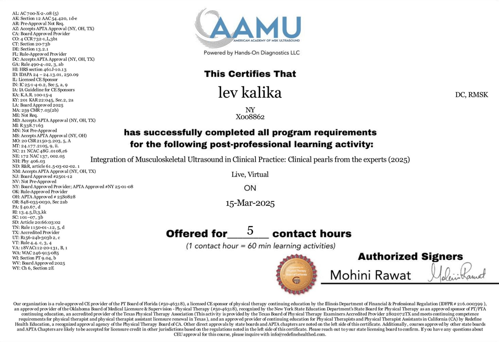
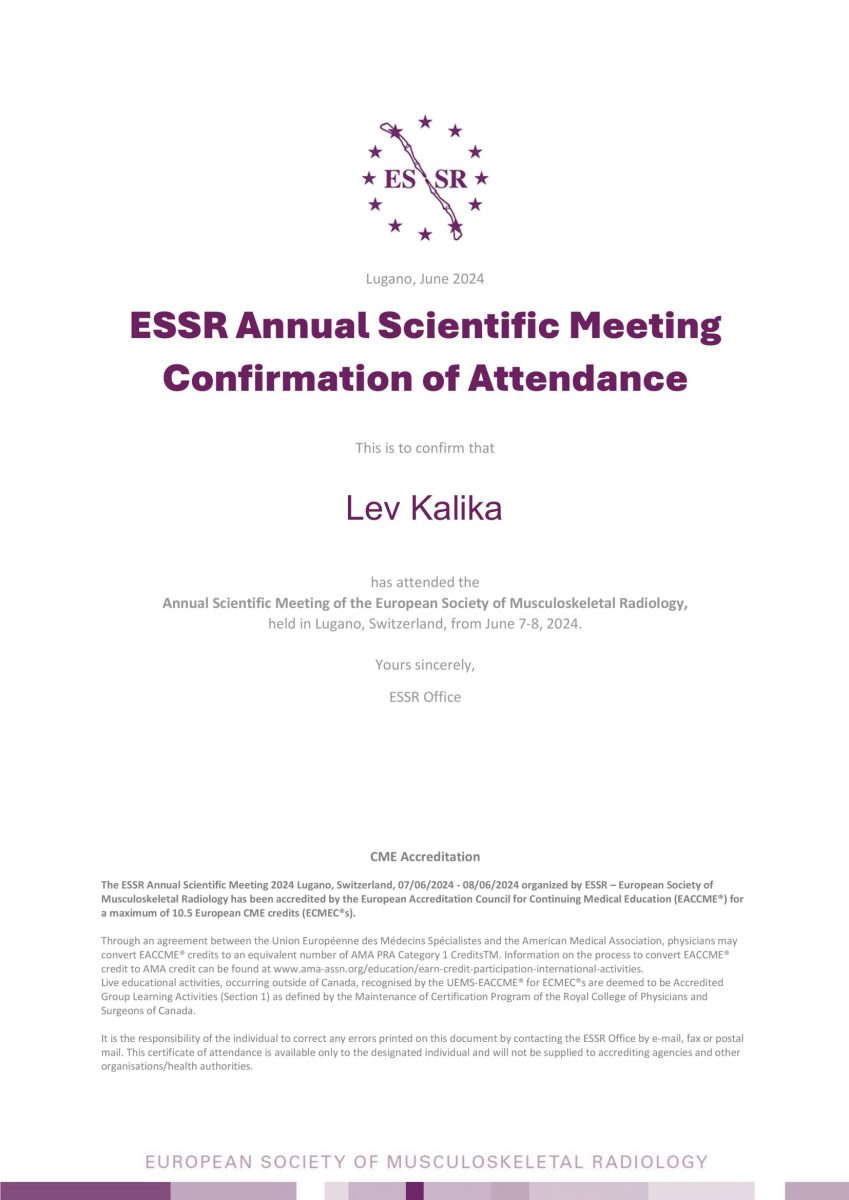
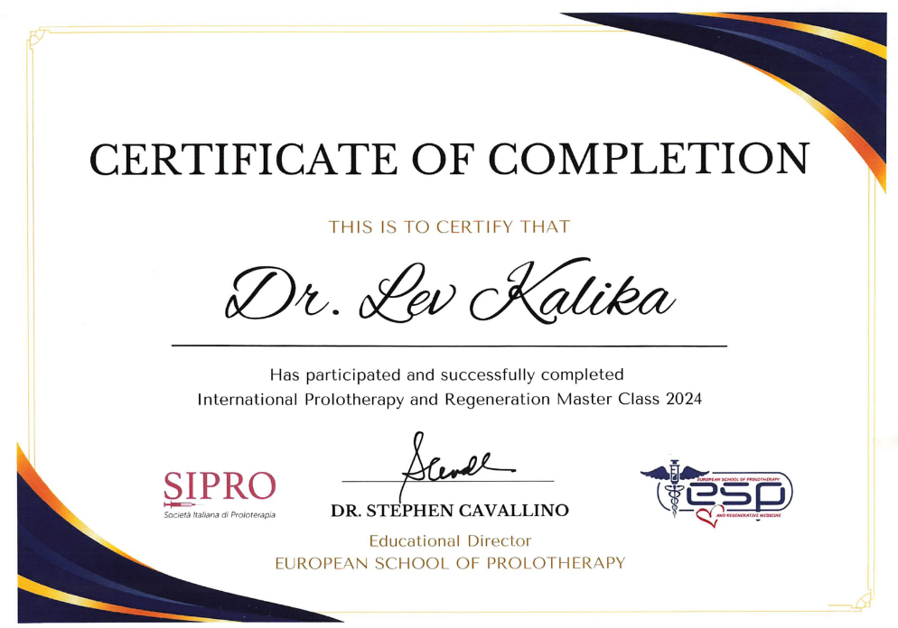
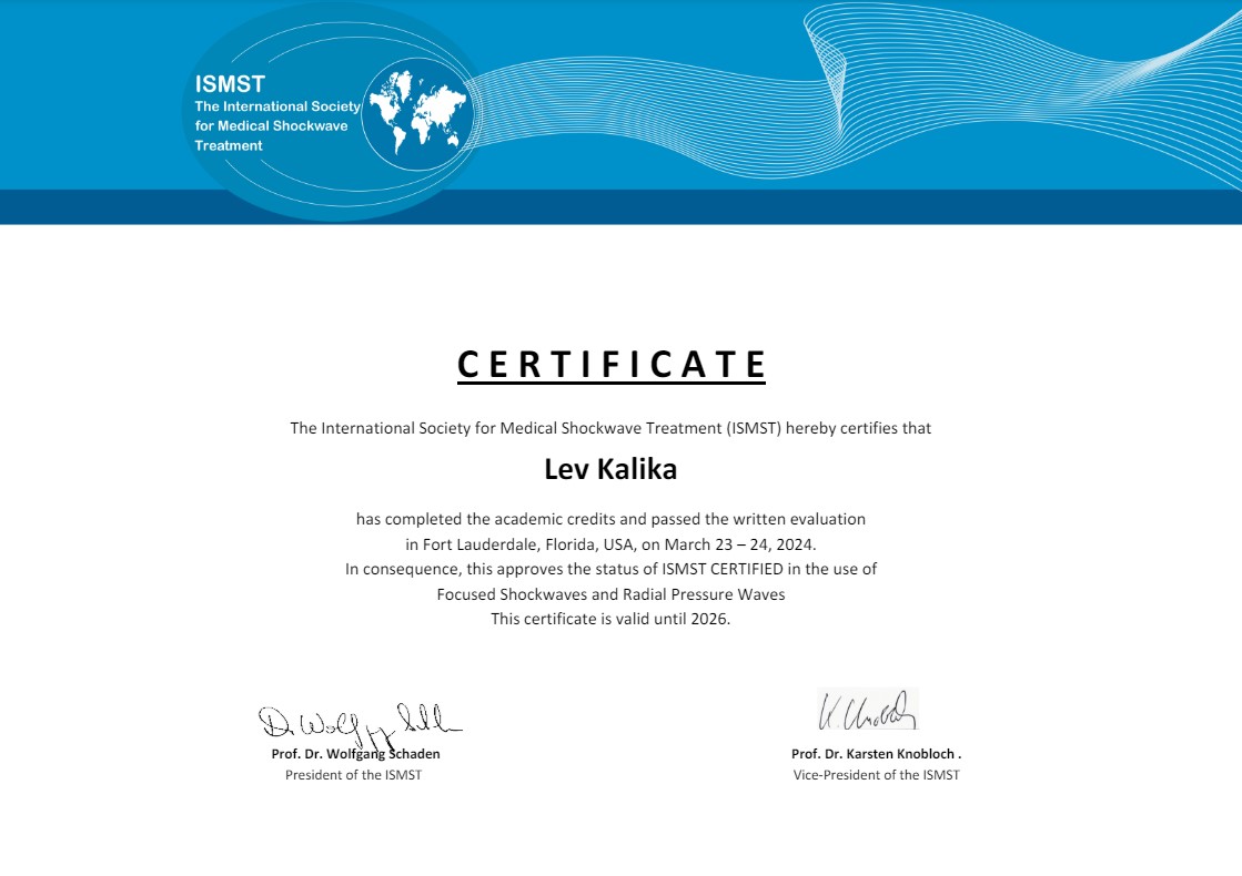
Dr. Lev Kalika is a world-recognized expert in musculoskeletal medicine. with 20+ years of clinical experience in diagnostic musculoskeletal ultrasonography, rehabilitative sports medicine and conservative orthopedics. In addition to operating his clinical practice in Manhattan, he regularly publishes peer-reviewed research on ultrasound-guided therapies and procedures. He serves as a peer reviewer for Springer Nature.
Dr. Kalika is an esteemed member of multiple professional organizations, including:

Below is a prime example of how ultrasound can take the guesswork out of diagnosis.

A bad physical therapy experience is one of the primary causes of unnecessary surgery

In this instance, an athlete was originally diagnosed with minor quadriceps muscle strain and was treated for four weeks, with unsatisfactory results. When he came to our clinic, the muscle was not healing, and the patients’ muscle tissue had already begun to atrophy.
Upon examination using MSUS, we discovered that he had a full muscle thickness tear that had been overlooked by his previous provider. To mitigate damage and promote healing, surgery should have been performed immediately after the injury occurred. Because of misdiagnosis and inappropriate treatment, the patient now has permanent damage that cannot be corrected.
The most important advantage of Ultrasound over MRI imaging is its ability to zero in on the symptomatic region and obtain imaging, with active participation and feedback from the patient. Using dynamic MSUS, we can see what happens when patients contract their muscles, something that cannot be done with MRI. From a diagnostic perspective, this interaction is invaluable.
Dynamic ultrasonography examination demonstrating
the full thickness tear and already occurring muscle atrophy
due to misdiagnosis and not referring the patient
to proper diagnostic workup
Demonstration of how very small muscle defect is made and revealed
to be a complete tear with muscle contraction
under diagnostic sonography (not possible with MRI)
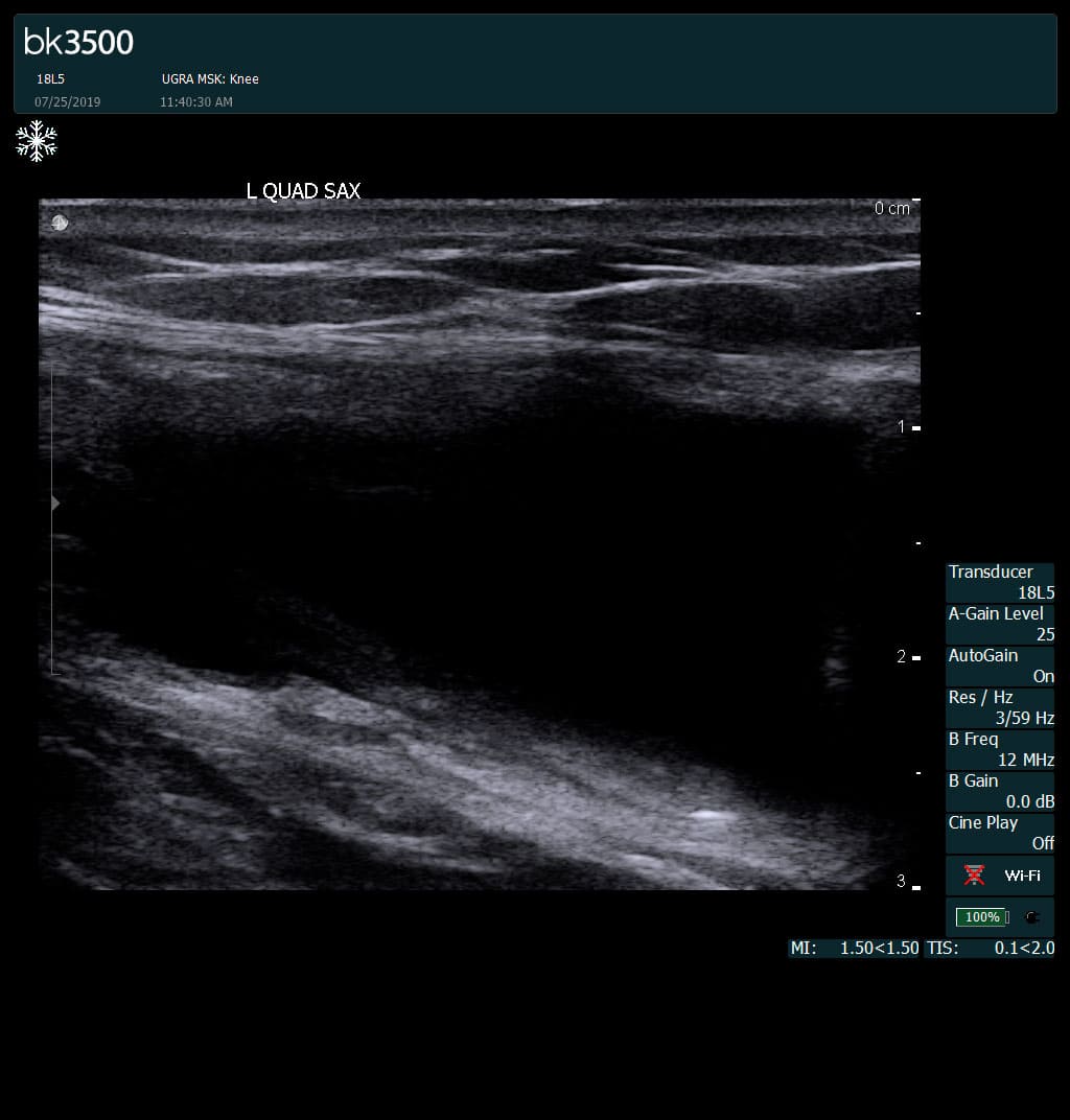
Complete tear of rectus femoris
with large hematoma (blood)
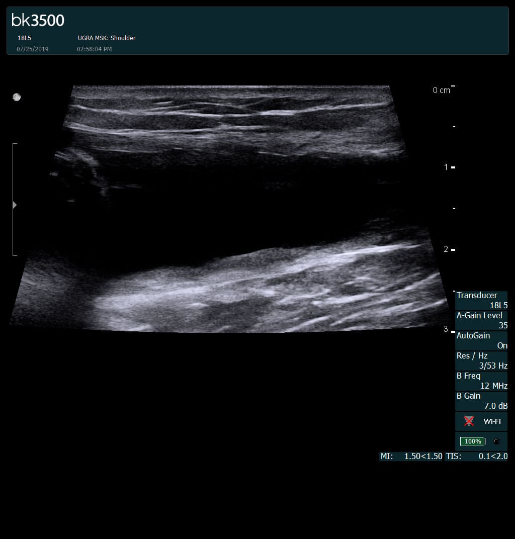
Separation of muscle ends due to tear elicited
on dynamic sonography examination


 Request Telehealth