MS Patients Find Balance Through C.A.R.E.N — Computer Assisted Rehabilitation Environment
A compromised sense of balance and resulting falls are key concerns for patients with multiple sclerosis (MS). Past balance-control strategies have relied on tasks coordinating motor skills with sensory input, Feldenkrais moves and stretching exercises.
However, a six-week pilot study published in the Journal of NeuroEngineering and Rehabilitation applied virtual reality (VR) gaming technology to improve balance and prevent debilitating falls.
Applying VR Balance Training to MS
MS is but one of a number of conditions associated with balance problems. C.A.R.E.N had already proved helpful for patients suffering from:
Orthopedic or musculoskeletal injuries or abnormalities
Dizziness or vertigo from vestibular or inner ear issues
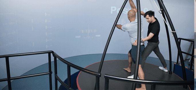
The system combines physical movement and motor abilities with cognitive perception and sensory skills, but it does so in a safe environment that is interactive and controlled yet also engaging and realistic.
The Computer Assisted Rehabilitation Environment
The C.A.R.E.N system uses a number of components to provide a realistic simulation with measurable results:
The patient stands on an electro hydraulic platform capable of movement: pitch, yaw and roll to coordinate with planned visuals.
A safety harness prevents falls but otherwise supports no body weight during exercise.
A wall-sized projection screen displays a virtual environment or scene that the patient must navigate from the platform. Meanwhile, the platform shifts, tilts and sways tours of the terrain directly ahead as the patient progresses through the scene.
Eighteen colorful ball targets heighten elements of challenge, engagement and skill. When a ball appears on the screen, patients have 5 seconds to move and balance.
Powering the entire experience, computer software allows either the participant or the physical therapist to increase or decrease challenge levels.
Evaluating Balance in MS Patients
Over six weeks, all study participants attended twelve 30-minute balance training sessions:
The VR group spent 30 minutes on the interactive computerized system.
The control group completed 10 minutes of stretching exercises and 20 minutes of balancing on a wobble board while catching balls tossed at varying directions and speeds.
At both the start and end of the six-week period, researchers recorded specific measurements through: Posturography. Tests were conducted with participants’ eyes first open and then closed for comparison. Treadmill data included:
Ellipse sway area
Center of pressure path (CoP) length
Sway rate
Average pressure distributions for both feet. The researchers also calculated and assigned asymmetry scores for the pressure distributions.
Clinical Balancing Tests. Participants completed a functional reach test, Berg Balance Test, Four Square Step Test and Falls Efficacy Scale International questionnaire.
How Effective Is Balance Training for MS Patients?
Improvements overall were significantly greater for the VR group. However, a few elements of the results offer deeper and perhaps promising insight:
Both the VR simulation group and the control group showed improved scores on clinical balancing tests and posturography data—particularly for CoP path length and sway rate.
Changes in CoP were especially significant, as past studies have correlated MS patients’ path length to falls as well as neurological damage.
Closed-eye posturography data demonstrated still greater improvement in the VR group over the control group versus comparisons of the two groups’ open-eye posturography data.
Virtual reality offers promising potential for improving the lives of MS patients through balance training. Too, computerized simulation systems increase confidence levels and compliance rates. For facilities able to access them— balance training is a worthwhile supplemental or complementary therapy intervention strategy of proven value.
In this instance, an athlete was originally diagnosed with minor quadriceps muscle strain and was treated for four weeks, with unsatisfactory results. When he came to our clinic, the muscle was not healing, and the patients’ muscle tissue had already begun to atrophy.
Upon examination using MSUS, we discovered that he had a full muscle thickness tear that had been overlooked by his previous provider. To mitigate damage and promote healing, surgery should have been performed immediately after the injury occurred. Because of misdiagnosis and inappropriate treatment, the patient now has permanent damage that cannot be corrected.
The most important advantage of Ultrasound over MRI imaging is its ability to zero in on the symptomatic region and obtain imaging, with active participation and feedback from the patient. Using dynamic MSUS, we can see what happens when patients contract their muscles, something that cannot be done with MRI. From a diagnostic perspective, this interaction is invaluable.
Dynamic ultrasonography examination demonstrating the full thickness tear and already occurring muscle atrophy due to misdiagnosis and not referring the patient to proper diagnostic workup
Demonstration of how very small muscle defect is made and revealed to be a complete tear with muscle contraction under diagnostic sonography (not possible with MRI)
Complete tear of rectus femoris with large hematoma (blood)
Separation of muscle ends due to tear elicited on dynamic sonography examination