Although there are many etiologies of low back pain and diversities within those origins, these are some common culprits of back pain:
Facet arthropathy
Herniated disc
Spondylosis
Stenosis
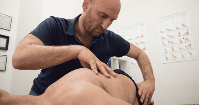
Also, trigger points are common sources, and trigger injections may control muscle spasms. The spasmodic movements are caused by chronic postures, which lead to stretch trauma in the weaker muscle groups. Therapies targeting these specific groups alone may actually be harmful by overworking the muscles. Micro-traumas have a combined effect that leads to completely eliminate the pain.
Spinal Mechanics
Increased or decreased curvature of the lumbar region can result in pain. Obesity, muscle hypertrophy and other issues may make diagnosis more difficult. Lordosis or outward curvature may be caused by the following:
Anterior pelvic tilt
Weak abdominal muscles
Shortened hip flexors
Posterior pelvic tilt may reduce lordosis. Since hip flexor shortening contributes greatly to pain, physicians should perform a strength test.
Lumbar Flexion
This flexion is a common daily activity while bending or standing. When bending, lumbar flexion should comprise 50 percent of the motion, and hip flexion should take over about halfway through the motion. When it does not, this leads to pain and possible muscle tearing. Extension tests that show a degree range above 30 indicate incorrect lumbar flexion and possible hip extension issues.
Lumbar Extension
With an anterior curve increase, this motion varies greatly in range. In most cases, damage is due to stenosis.
Rotational Motion
Lower spine rotation should be about 13 degrees when the applicable segments contribute correctly. Also, rotational restriction is completed by the thoracic spine. If symmetry is lacking between the lengths and strengths of the abdominal muscles, mobility is limited. As a result, there is a difference in muscle mass. The outcome is a postural change that leads to pain. Strengthening the weakened muscles is an important corrective component.
Lateral Flexion
Most mobility in this area is in the lower thoracic spine. Lateral flexion rotates in the direction of the curve convexity. Since segmental and lateral movements work together, impairment of one will affect the other. Limited side bending capabilities and pain are the results.
Since there are so many muscle groups and regions affecting low back pain, it is important together. Treating the pain with back strengthening alone will not suffice. There must also be abdominal strengthening and opposing hip flexor lengthening.
In this instance, an athlete was originally diagnosed with minor quadriceps muscle strain and was treated for four weeks, with unsatisfactory results. When he came to our clinic, the muscle was not healing, and the patients’ muscle tissue had already begun to atrophy.
Upon examination using MSUS, we discovered that he had a full muscle thickness tear that had been overlooked by his previous provider. To mitigate damage and promote healing, surgery should have been performed immediately after the injury occurred. Because of misdiagnosis and inappropriate treatment, the patient now has permanent damage that cannot be corrected.
The most important advantage of Ultrasound over MRI imaging is its ability to zero in on the symptomatic region and obtain imaging, with active participation and feedback from the patient. Using dynamic MSUS, we can see what happens when patients contract their muscles, something that cannot be done with MRI. From a diagnostic perspective, this interaction is invaluable.
Dynamic ultrasonography examination demonstrating the full thickness tear and already occurring muscle atrophy due to misdiagnosis and not referring the patient to proper diagnostic workup
Demonstration of how very small muscle defect is made and revealed to be a complete tear with muscle contraction under diagnostic sonography (not possible with MRI)
Complete tear of rectus femoris with large hematoma (blood)
Separation of muscle ends due to tear elicited on dynamic sonography examination