August 14, 2023
Peroneal Tendinopathy, also called peroneal tendinitis, is a relatively rare but painful tendon injury that results from damage and degeneration of a peroneal tendon in the foot. The condition is most often associated with running, but basketball players, dancers and other athletes in sports that involve jumping and landing are also prone to the condition. PT is characterized by pain that begins near the ankle at the outside of the foot, and runs up the outer lower leg.
Each foot has two peroneal tendons, longus and the brevis, and tendinopathy can occur in either or both tendons. The tendons connect the peroneus longus and the peroneus brevis muscles toes.
Damage to the peroneal tendons often occurs from overuse of the muscles during daily running, sports practice, rehearsals and performance. Injuries are most common in people with high arches, which places greater stress on the peroneal tendons. High arches are considered an asset in dancers, placing them at greater risk. Faster running speeds also increase peroneal tendon workload, and peroneal tendinopathy often accompanies an ankle sprain. The condition worsens with continued activity and improves with rest.
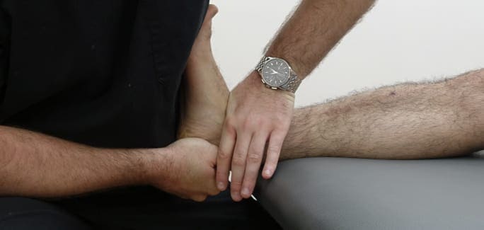
To differentiate peroneal tendinopathy from other sources of foot pain, a through health histo visual the internal structures of your foot and ankle in real time, with feedback from the athlete facilitating an accurate diagnosis.
Other diagnostic tests and assessments may include:
Once the therapist has confirmed an injury to the peroneal tendon, an individualized treatment plan will be designed. Treatment may include:
Treatment at NYDNR is holistic, individualized and multi-modal. If you are experiencing pain in your foot and ankle, failure today, and see why we are the foremost clinic for foot and ankle rehabilitation in NYC.

Verified Expert Profiles
Dr. Lev Kalika is a world-recognized expert in musculoskeletal medicine. with 20+ years of clinical experience in diagnostic musculoskeletal ultrasonography, rehabilitative sports medicine and conservative orthopedics. In addition to operating his clinical practice in Manhattan, he regularly publishes peer-reviewed research on ultrasound-guided therapies and procedures. He serves as a peer reviewer for Springer Nature.
Dr. Kalika is an esteemed member of multiple professional organizations, including:
Below is a prime example of how ultrasound can take the guesswork out of diagnosis.

A bad physical therapy experience is one of the primary causes of unnecessary surgery
In this instance, an athlete was originally diagnosed with minor quadriceps muscle strain and was treated for four weeks, with unsatisfactory results. When he came to our clinic, the muscle was not healing, and the patients’ muscle tissue had already begun to atrophy.
Upon examination using MSUS, we discovered that he had a full muscle thickness tear that had been overlooked by his previous provider. To mitigate damage and promote healing, surgery should have been performed immediately after the injury occurred. Because of misdiagnosis and inappropriate treatment, the patient now has permanent damage that cannot be corrected.
The most important advantage of Ultrasound over MRI imaging is its ability to zero in on the symptomatic region and obtain imaging, with active participation and feedback from the patient. Using dynamic MSUS, we can see what happens when patients contract their muscles, something that cannot be done with MRI. From a diagnostic perspective, this interaction is invaluable.
Dynamic ultrasonography examination demonstrating
the full thickness tear and already occurring muscle atrophy
due to misdiagnosis and not referring the patient
to proper diagnostic workup
Demonstration of how very small muscle defect is made and revealed
to be a complete tear with muscle contraction
under diagnostic sonography (not possible with MRI)

Complete tear of rectus femoris
with large hematoma (blood)

Separation of muscle ends due to tear elicited
on dynamic sonography examination


