HomeBlogPhysical Therapy Provides Cost-Saving, Effective Treatment of Low Back Pain
Physical Therapy Provides Cost-Saving, Effective Treatment of Low Back Pain
When a patient visits a docto the normal aging process.
A Pain in the Wallet
An MRI may provide peace of mind, but new research shows that in most cases the imaging procedure only adds a pain in the wallet on to a study led by a professor in the field of physical therapy.
An MRI often shows many changes and abnormalities of the back and spine, but most of these findings are benign. The patient then becomes concerned and puts a large amount of pressure on docto cure his or her condition. This results in the patient being referred for surgery, injections and specialist visits. These are costs that add up quickly.
Patients Seek Treatment in Absence of Pain
Patients often ask for these services even in the absence of pain, discomfort or sympto the point where it causes pain or discomfort. The study found that when patients get this diagnosis from an MRI, they usually ask for treatment.
The Costly Burden of Back Pain
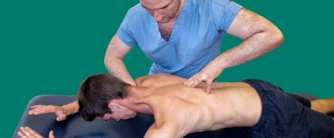
Pain in the lower back is one of the most prevalent and disabling health problems worldwide. People spend more time out of work due to back pain than other more serious conditions, such as cancer, traffic injuries and childbirth.
According to the billions of dollars worth of lost hours each year and it becomes obvious that back pain is a costly epidemic. However, we found that there are more cost-effective methods for treating back pain.
A Less Expensive, More Effective Alternative
A better option for most patients complaining of low back pain is tor for their back pain. Patients who received an MRI first spent $6,664 during the same amount of time.
When low back pain patients go to reduce it on their own. The study speculated that patients who chose this option were more engaged with their therapy and more motivated for success in their own recovery, while patients who went with surgery or injections expected a quick fix.
Beginning treatment with physical therapy rather than an MRI does not rule out the option of having an MRI if needed. When a patient has specific symptor said that advanced imaging has its place, but that place is not in the beginning of treatment for the majority of patients.
Convincing Patients to Try Physical Therapy First
Many people are used to check for cavities or broken bones, but many other conditions including low back pain are better assessed through other means. The study observed that patients often find it unacceptable when imaging is denied or discouraged in favor of physical therapy or other treatments first.
Physicians can help patients by explaining the monetary savings of trying physical therapy first. Using physical therapy as the first-line treatment instead of an MRI also leads to the extent that they no longer seek other treatments. Physicians can also point out that there is no harm in trying physical therapy and that other treatments can always be tried later if the patient does not improve.
In this instance, an athlete was originally diagnosed with minor quadriceps muscle strain and was treated for four weeks, with unsatisfactory results. When he came to our clinic, the muscle was not healing, and the patients’ muscle tissue had already begun to atrophy.
Upon examination using MSUS, we discovered that he had a full muscle thickness tear that had been overlooked by his previous provider. To mitigate damage and promote healing, surgery should have been performed immediately after the injury occurred. Because of misdiagnosis and inappropriate treatment, the patient now has permanent damage that cannot be corrected.
The most important advantage of Ultrasound over MRI imaging is its ability to zero in on the symptomatic region and obtain imaging, with active participation and feedback from the patient. Using dynamic MSUS, we can see what happens when patients contract their muscles, something that cannot be done with MRI. From a diagnostic perspective, this interaction is invaluable.
Dynamic ultrasonography examination demonstrating the full thickness tear and already occurring muscle atrophy due to misdiagnosis and not referring the patient to proper diagnostic workup
Demonstration of how very small muscle defect is made and revealed to be a complete tear with muscle contraction under diagnostic sonography (not possible with MRI)
Complete tear of rectus femoris with large hematoma (blood)
Separation of muscle ends due to tear elicited on dynamic sonography examination