August 14, 2023
Each year, between 10 and 13 percent of reported sports-related injuries involve groin pain. Some of these people will be diagnosed with athletic pubalgia, which is also known as a sports hernia. These are injuries that may happen to continue participating in his or her chosen sport.
When a patient reports groin pain, doctors should first check for other conditions that can cause the pain. There are a number of differential diagnoses, including:
There are several other possible causes of pain in the groin. The differential diagnoses should be eliminated as causing the pain first.
People who are suffering from athletic pubalgia often report pain in their groins and lower abdomens that may radiate down to play despite the pain.
When a sports hernia is suspected, a docto confirm the diagnosis.
Sports hernias will generally need to finish out the season.
After surgery, rehabilitation will involve several things. Athletes will be able to their sports at about six weeks after their surgeries.
Groin pain is a fairly common type of injury experienced by people who play certain sports. When a sports hernia is suspected, it is important that it is properly diagnosed and treated so that it doesn’t prevent the person from being able to his or her sport.

Verified Expert Profiles
Dr. Lev Kalika is a world-recognized expert in musculoskeletal medicine. with 20+ years of clinical experience in diagnostic musculoskeletal ultrasonography, rehabilitative sports medicine and conservative orthopedics. In addition to operating his clinical practice in Manhattan, he regularly publishes peer-reviewed research on ultrasound-guided therapies and procedures. He serves as a peer reviewer for Springer Nature.
Dr. Kalika is an esteemed member of multiple professional organizations, including:
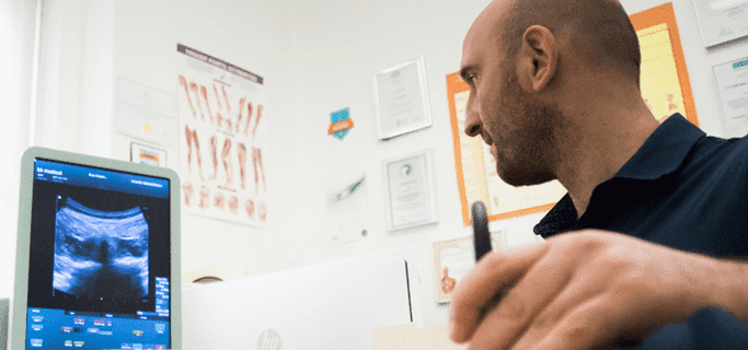
Below is a prime example of how ultrasound can take the guesswork out of diagnosis.

A bad physical therapy experience is one of the primary causes of unnecessary surgery
In this instance, an athlete was originally diagnosed with minor quadriceps muscle strain and was treated for four weeks, with unsatisfactory results. When he came to our clinic, the muscle was not healing, and the patients’ muscle tissue had already begun to atrophy.
Upon examination using MSUS, we discovered that he had a full muscle thickness tear that had been overlooked by his previous provider. To mitigate damage and promote healing, surgery should have been performed immediately after the injury occurred. Because of misdiagnosis and inappropriate treatment, the patient now has permanent damage that cannot be corrected.
The most important advantage of Ultrasound over MRI imaging is its ability to zero in on the symptomatic region and obtain imaging, with active participation and feedback from the patient. Using dynamic MSUS, we can see what happens when patients contract their muscles, something that cannot be done with MRI. From a diagnostic perspective, this interaction is invaluable.
Dynamic ultrasonography examination demonstrating
the full thickness tear and already occurring muscle atrophy
due to misdiagnosis and not referring the patient
to proper diagnostic workup
Demonstration of how very small muscle defect is made and revealed
to be a complete tear with muscle contraction
under diagnostic sonography (not possible with MRI)

Complete tear of rectus femoris
with large hematoma (blood)

Separation of muscle ends due to tear elicited
on dynamic sonography examination


