HomeBlogRecognizing and Treating Plantar Fasciitis
Recognizing and Treating Plantar Fasciitis
Foot and heel pain can really slow you down, keeping you from activities you love and making every step a chore. In many cases, pain that begins in your heel is caused by plantar fasciitis, an inflammatoes. In some cases, tiny micro tears of the plantar fascia tissue aggravate the condition, increasing pain and prolonging healing time.
Many factors contribute the growing number of cases of plantar fasciitis each year:
• Increased body weight • Walking and standing on hard surfaces • Physical inactivity • Excessive forefoot sports • Non supportive footwear • Increased longevity
Plantar fasciitis is a type of overuse injury that grows worse over time unless treated.
Symptoms include:
• Heel and foot pain first thing in the morning • Aching and throbbing in the evening • Pain when standing after long periods of sitting • Severe pain after sports or exercise
Phases of Plantar Fasciitis
Plantar fasciitis typically progresses through three phases:
Phase I: Pain begins as dull heel pain that may feel like a sto four weeks’ time. Pain intensifies with weight bearing activity and is at its worst when your feet hit the floor first thing in the morning. Ice, rest, stretching and supportive footwear may help relieve pain.
Phase II: Pain intensifies over six to cortisone shots and considering surgery.
Phase III: Pain begins to exercise can make matters worse.
Left untreated, pain from plantar fasciitis can last from two to five years. Even after recovery, the condition may return once normal activities are resumed.
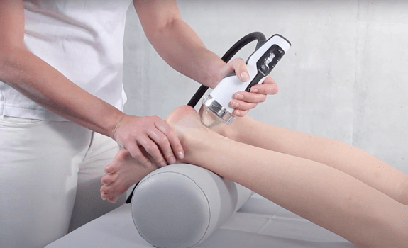
Plantar Fasciitis Treatment at NYDNRehab
The sports medicine professionals at NYDNRehab use diagnostic ultrasonography to disrupt the cycle of pain and inflammation.
You do not have to recovery.
In this instance, an athlete was originally diagnosed with minor quadriceps muscle strain and was treated for four weeks, with unsatisfactory results. When he came to our clinic, the muscle was not healing, and the patients’ muscle tissue had already begun to atrophy.
Upon examination using MSUS, we discovered that he had a full muscle thickness tear that had been overlooked by his previous provider. To mitigate damage and promote healing, surgery should have been performed immediately after the injury occurred. Because of misdiagnosis and inappropriate treatment, the patient now has permanent damage that cannot be corrected.
The most important advantage of Ultrasound over MRI imaging is its ability to zero in on the symptomatic region and obtain imaging, with active participation and feedback from the patient. Using dynamic MSUS, we can see what happens when patients contract their muscles, something that cannot be done with MRI. From a diagnostic perspective, this interaction is invaluable.
Dynamic ultrasonography examination demonstrating the full thickness tear and already occurring muscle atrophy due to misdiagnosis and not referring the patient to proper diagnostic workup
Demonstration of how very small muscle defect is made and revealed to be a complete tear with muscle contraction under diagnostic sonography (not possible with MRI)
Complete tear of rectus femoris with large hematoma (blood)
Separation of muscle ends due to tear elicited on dynamic sonography examination