HomeBlogRunning Footwear: Safety Gear or Setup for Injury?
Running Footwear: Safety Gear or Setup for Injury?
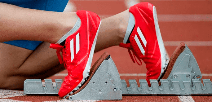
Since the advent of the running craze in the 1970s, the athletic shoe industry has been perpetually evolving, adapting new technologies and materials to maximize absorption of shock, gait stabilization, arch support.
Recently, shoe makers have even added wearable tech to the selection. Yet despite those apparent style and functionality alterations, the basics of athletic shoe structure remain:
corrective control of movement, balance, to trauma.
Analysis Challenges shoe design background based on how sport shoes producers categorize shoe functionality in terms of the human fascia shape, determined as low, medium or high. There is a concept to compensate for variance in running principles by “correcting” a runner’s gait.
An actual study held by Knapik et al. used conscripts to people in accordance with their plantar shape.
The trainees taking part in the research were male and female marines, whose feet were evaluated based on how much plantar surface came in contact with a specialized platform. Trained evaluato two groups. The key group of marine freshmen (400+ men and 250+ women) got the same shoe style.
The other group was given the shoes based on their foot shape: over 400 men and over 300 women. A group of medical assistants were tracking the trauma during twelve week’s time. No difference in the amount of injury between those two groups was found. This way the research group concluded that there is no connection of the shoe type or plantar shape to trauma occurrence.
An even larger research, held by same research team in 2014 corroborated the findings. Comfort Trumps Shoe Design An even more recent study conducted in 2017 evaluated running kinematics of 35 heel-toe runners in all three shoe types, and during barefoot running. The study’s authors concluded that most runners maintain the preferred movement path, no matter the type of the shoes they’re wearing.
The result of this research suggests that the shoes designed for specific fascia types have little to no impact on essential gait mechanics.
It would appear runners and other athletes should select shoes according to preferred comfort levels rather than category.
Running Gait Analysis and Retraining at NYDNRehab
Shoes made for running may not live up tor cuff tear, myofascial pain, sciatica pain, meniscus tears.
The highly trained professionals of our clinic base their diagnosis conclusions and treatment prescriptions exclusively on objective knowledge of medical science.
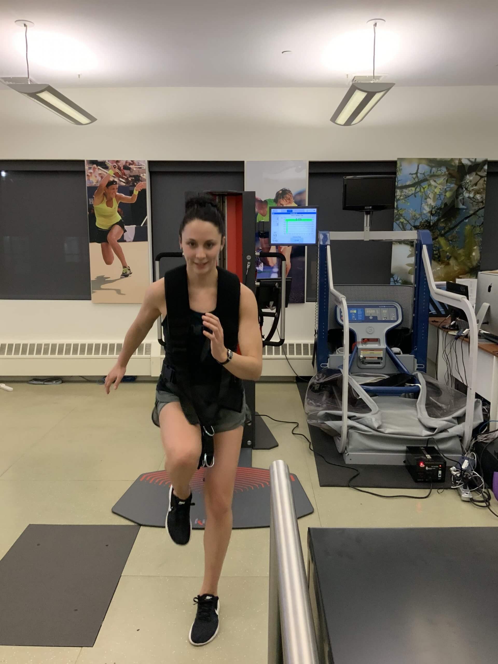
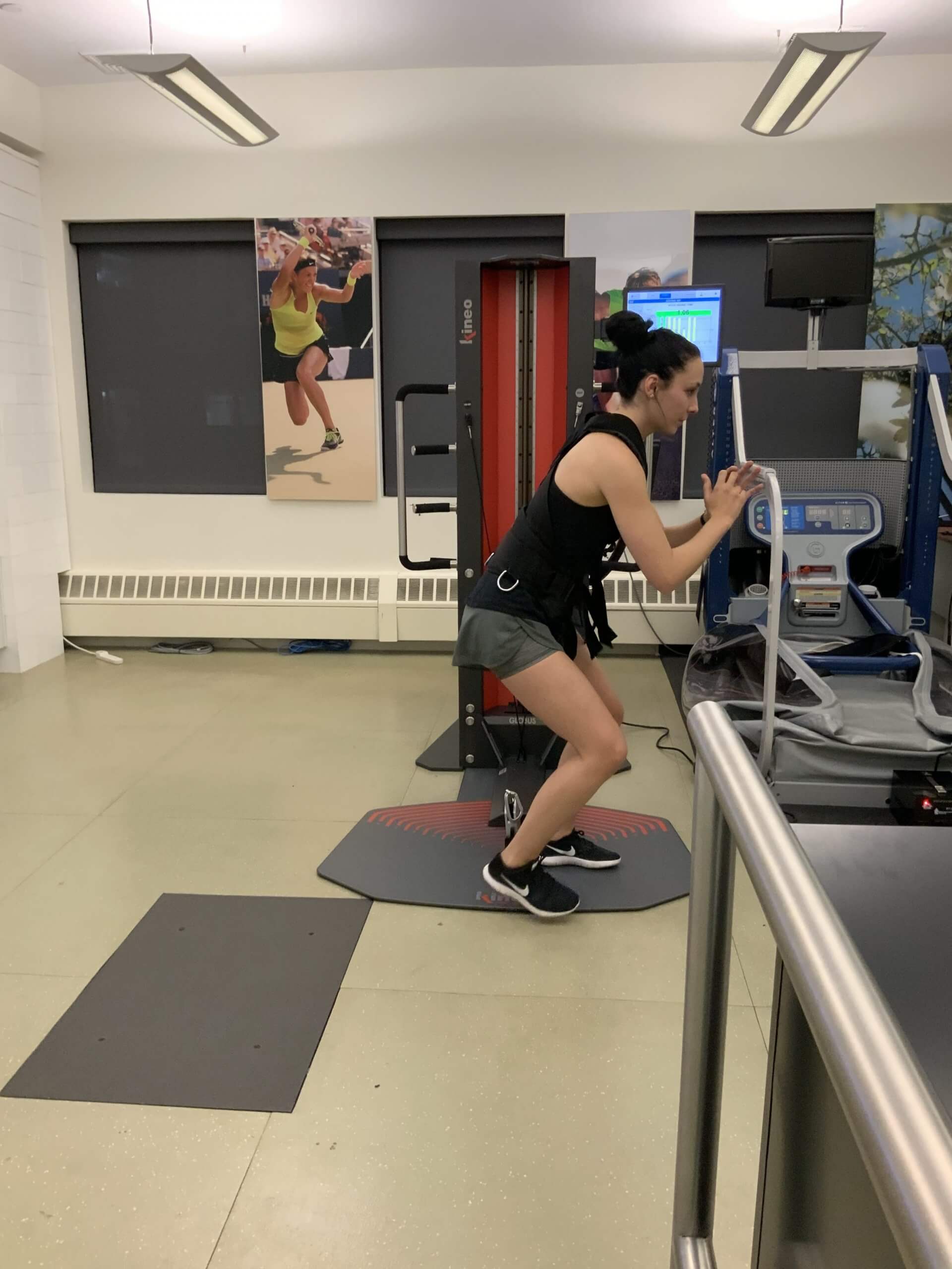
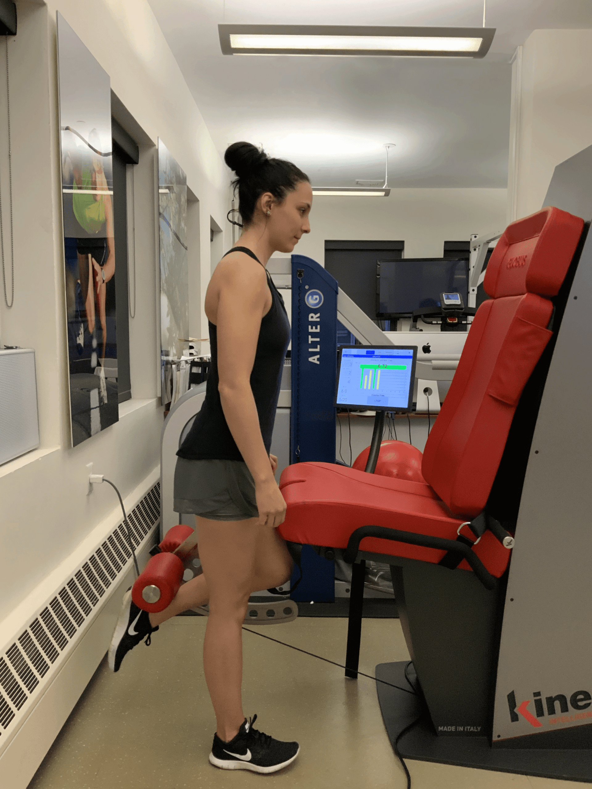
Reactive Neuromuscular Training on Kineo
Kineo – the most versatile muscle testing using artificial intelegence
Kineo – the most versatile muscle testing using artificial intelegence
Kineo – the most versatile muscle testing using artificial intelegence
In this instance, an athlete was originally diagnosed with minor quadriceps muscle strain and was treated for four weeks, with unsatisfactory results. When he came to our clinic, the muscle was not healing, and the patients’ muscle tissue had already begun to atrophy.
Upon examination using MSUS, we discovered that he had a full muscle thickness tear that had been overlooked by his previous provider. To mitigate damage and promote healing, surgery should have been performed immediately after the injury occurred. Because of misdiagnosis and inappropriate treatment, the patient now has permanent damage that cannot be corrected.
The most important advantage of Ultrasound over MRI imaging is its ability to zero in on the symptomatic region and obtain imaging, with active participation and feedback from the patient. Using dynamic MSUS, we can see what happens when patients contract their muscles, something that cannot be done with MRI. From a diagnostic perspective, this interaction is invaluable.
Dynamic ultrasonography examination demonstrating the full thickness tear and already occurring muscle atrophy due to misdiagnosis and not referring the patient to proper diagnostic workup
Demonstration of how very small muscle defect is made and revealed to be a complete tear with muscle contraction under diagnostic sonography (not possible with MRI)
Complete tear of rectus femoris with large hematoma (blood)
Separation of muscle ends due to tear elicited on dynamic sonography examination