Treatment of Running Injuries Symposium

I just got back from the first symposium on the world of running medicine and running injury treatment. It was an unusual symposium. Most tors and researchers presenting the studies with every detail of how the study was conducted and etc..

Sometimes you notice some listeners fall asleep. This one was quite different. There were only four presenters. Three of whom are tor of National Institute of running at Harvard University. Irene has also published over a hundred research studies and articles in scientific journals. She is considered the world’s authority on running retraining, foot biomechanics and barefoot running. Brian Hidershide is a young researcher from the University of Wisconsin who is considered a world’s expert on treatment and management of running injuries by manipulation of spatio-temporal parameters.

This symposium was designed in such a way that each of the presenters had to find out what other methods of running injury treatment will be discussed.

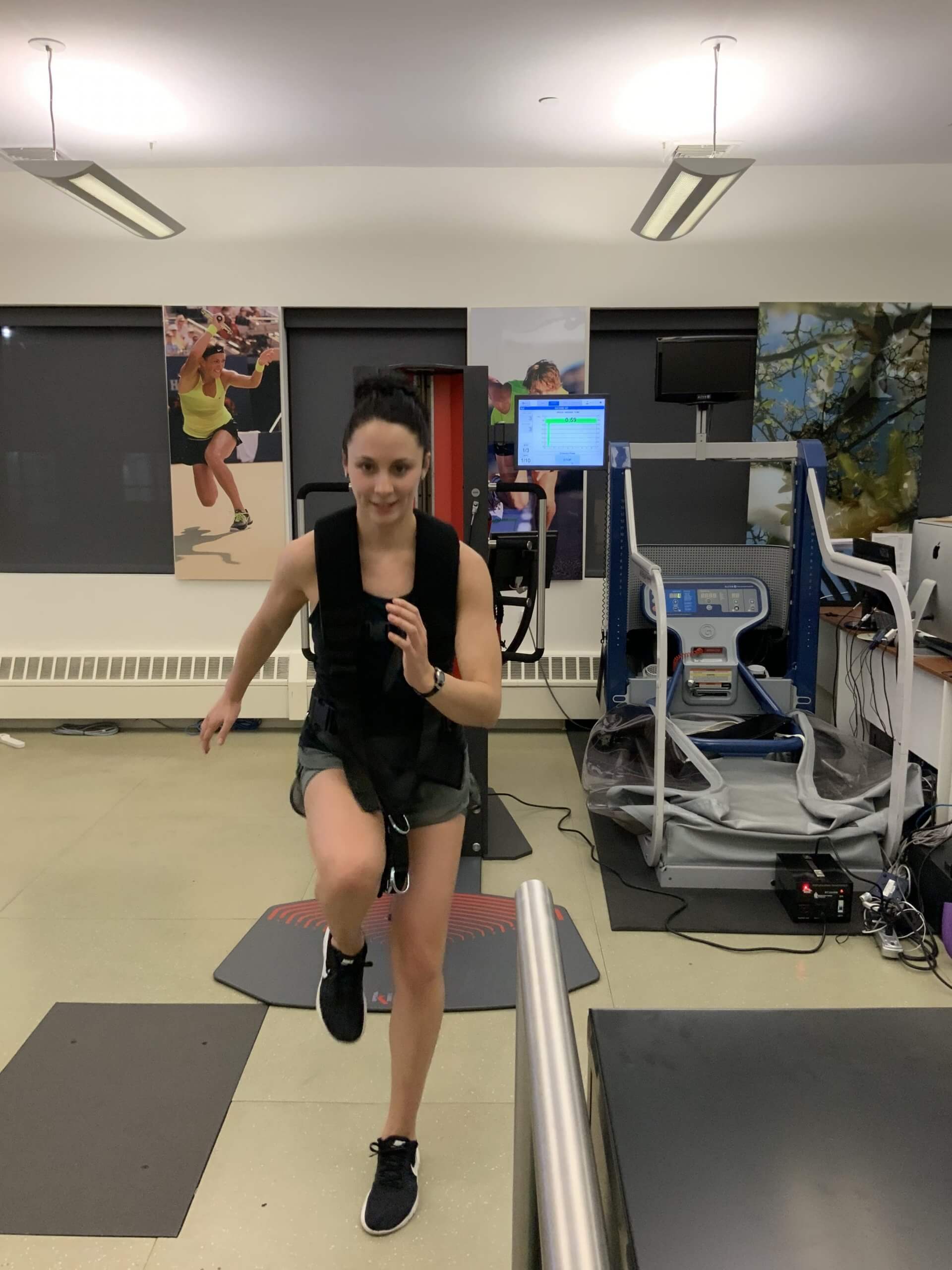
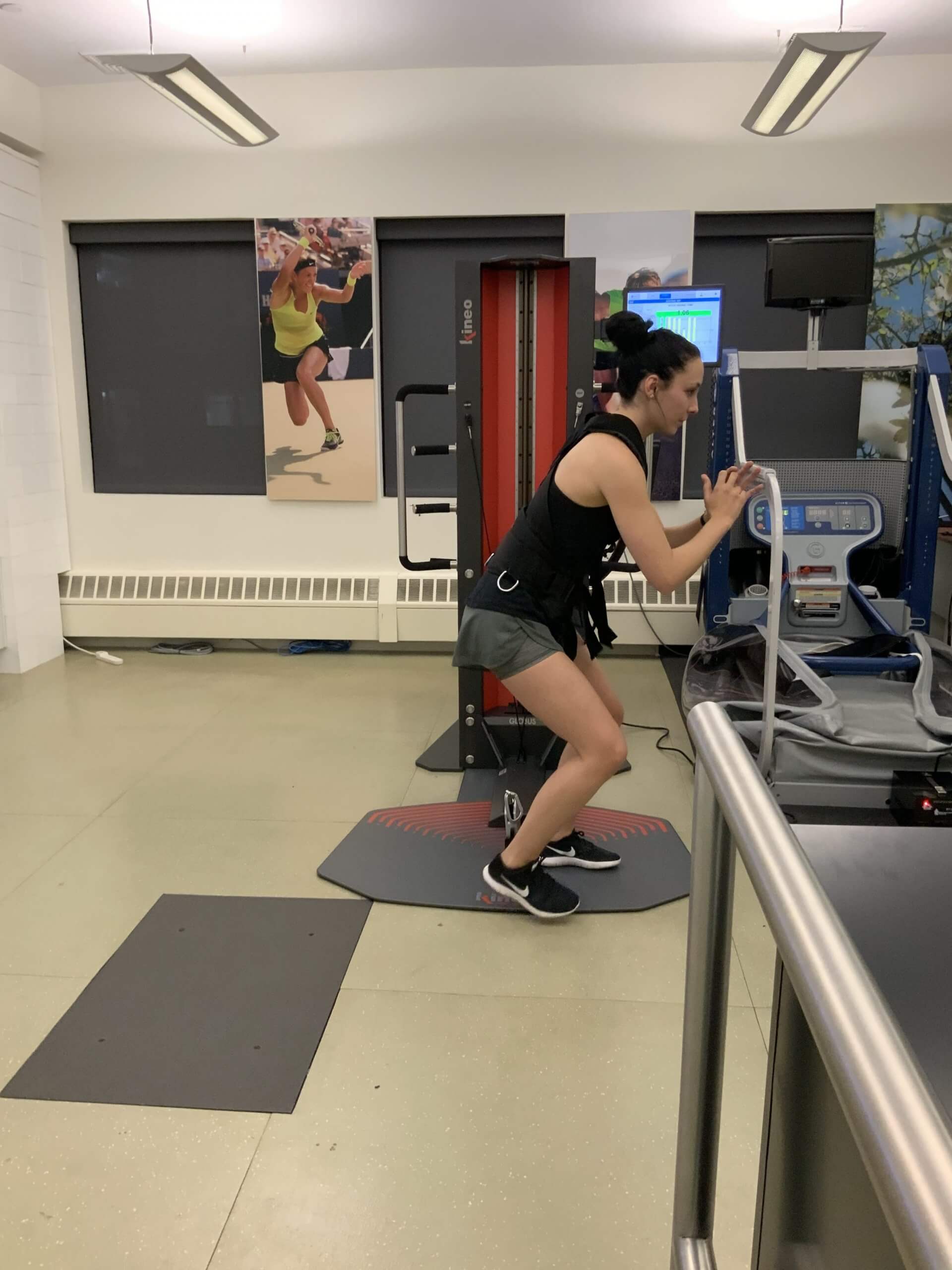
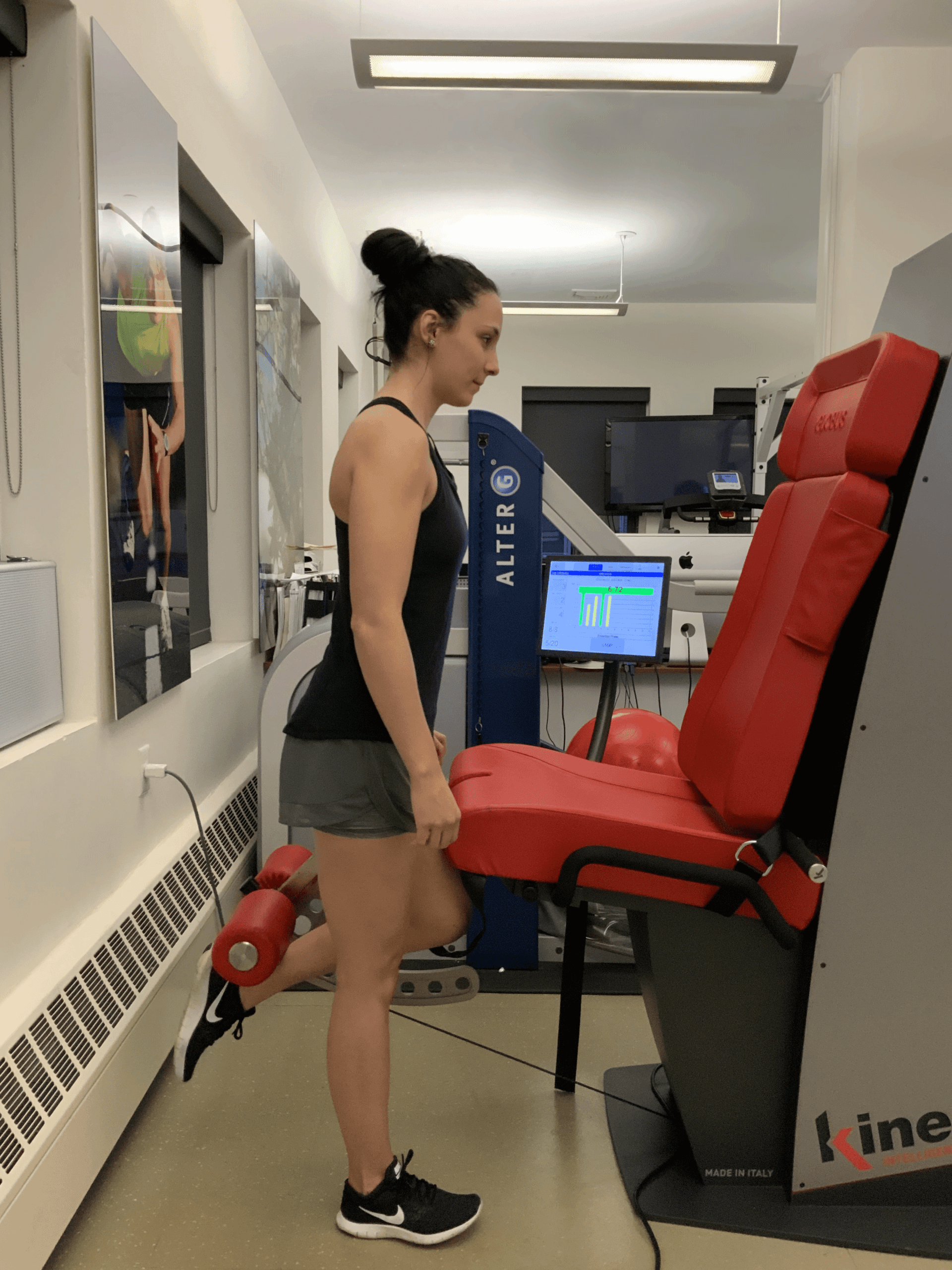
Reactive Neuromuscular Training on Kineo

pic

Kineo – the most versatile muscle testing using artificial intelegence

pic

Kineo – the most versatile muscle testing using artificial intelegence

pic

Kineo – the most versatile muscle testing using artificial intelegence

bg

In this instance, an athlete was originally diagnosed with minor quadriceps muscle strain and was treated for four weeks, with unsatisfactory results. When he came to our clinic, the muscle was not healing, and the patients’ muscle tissue had already begun to atrophy.

Upon examination using MSUS, we discovered that he had a full muscle thickness tear that had been overlooked by his previous provider. To mitigate damage and promote healing, surgery should have been performed immediately after the injury occurred. Because of misdiagnosis and inappropriate treatment, the patient now has permanent damage that cannot be corrected.

The most important advantage of Ultrasound over MRI imaging is its ability to zero in on the symptomatic region and obtain imaging, with active participation and feedback from the patient. Using dynamic MSUS, we can see what happens when patients contract their muscles, something that cannot be done with MRI. From a diagnostic perspective, this interaction is invaluable.

Dynamic ultrasonography examination demonstrating
the full thickness tear and already occurring muscle atrophy
due to misdiagnosis and not referring the patient
to proper diagnostic workup

Demonstration of how very small muscle defect is made and revealed
to be a complete tear with muscle contraction
under diagnostic sonography (not possible with MRI)

image

Complete tear of rectus femoris
with large hematoma (blood)

image

Separation of muscle ends due to tear elicited
on dynamic sonography examination

bg
Buy now 3D Gait
Payment Success
Request Telehealth Request Telehealth Request in office visit Book now