November 4, 2024
If a patient is suffering from calcific shoulder tendonitis, the condition may cause severe pain, limit the individual’s range of motion, increase the production of inflammato the production of new tissue.
Numerous studies have suggested that extracorporeal shockwave therapy can increase the amount of blood that will reach the bones and the connective tissue, and the treatment may boost the level of oxygen that positively affects the tendons and increase the amount of vitamins that the joint absorbs. Furthermore, the treatments may cause the body to produce additional capillaries, which are small blood vessels that may accelerate recovery and hinder the formation of additional deposits of calcium.
When specialists perform the procedures, the patient will not experience pain or discomfort, and unlike many alternative therapies, the cutting-edge technology will never cause side effects. Moreover, the treatments will not require the patient to undergo a recovery period.
The therapy may improve the thickness of the connective tissue by prompting the cells to one another and may slightly extend the lifespans of some cells.
Multiple analyses have shown that the newly regenerated cells are able to improve the strength of the shoulder. Likewise, the cells can swiftly reduce tenderness that calcific shoulder tendonitis may cause.
Decreasing Pain
The condition is commonly precipitated by repetitive movements that slowly wear down the rotator cuff’s tissue, and eventually, the deposits of calcium will cause pain and inflammation by rubbing against the tendons when the shoulder moves. The treatment can decrease the production of cyclooxygenase, which may precipitate oxidation, augment the body’s levels of prostaglandins and reduce the density of the tissue.
According to 82 percent of calcific deposits.
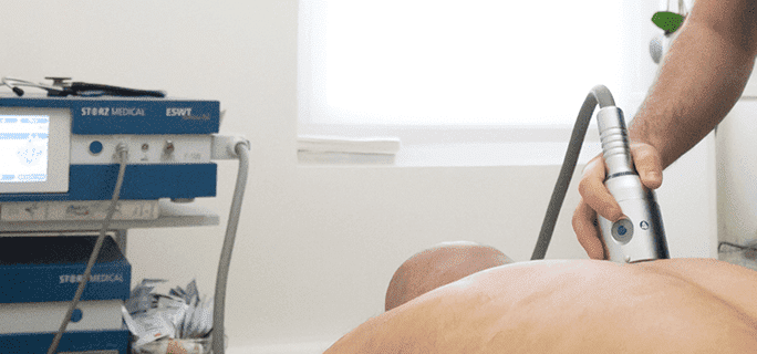
During each appointment, the specialists will utilize a device with a cutting-edge coil that generates controlled bursts of energy. If an injured athlete chooses extracorporeal shockwave therapy, the patient will generally have to undergo at least three sessions, and a clinic may provide one treatment per week. Sometimes, each session will have a duration of less than 20 minutes.
Various analyses have indicated that an individual will notice a decrease in pain within three weeks of the first treatment. Additionally, multiple studies showed that the therapy’s effects are typically evident for more than one year.

Verified Expert Profiles
Dr. Lev Kalika is a world-recognized expert in musculoskeletal medicine. with 20+ years of clinical experience in diagnostic musculoskeletal ultrasonography, rehabilitative sports medicine and conservative orthopedics. In addition to operating his clinical practice in Manhattan, he regularly publishes peer-reviewed research on ultrasound-guided therapies and procedures. He serves as a peer reviewer for Springer Nature.
Dr. Kalika is an esteemed member of multiple professional organizations, including:
Below is a prime example of how ultrasound can take the guesswork out of diagnosis.

A bad physical therapy experience is one of the primary causes of unnecessary surgery
In this instance, an athlete was originally diagnosed with minor quadriceps muscle strain and was treated for four weeks, with unsatisfactory results. When he came to our clinic, the muscle was not healing, and the patients’ muscle tissue had already begun to atrophy.
Upon examination using MSUS, we discovered that he had a full muscle thickness tear that had been overlooked by his previous provider. To mitigate damage and promote healing, surgery should have been performed immediately after the injury occurred. Because of misdiagnosis and inappropriate treatment, the patient now has permanent damage that cannot be corrected.
The most important advantage of Ultrasound over MRI imaging is its ability to zero in on the symptomatic region and obtain imaging, with active participation and feedback from the patient. Using dynamic MSUS, we can see what happens when patients contract their muscles, something that cannot be done with MRI. From a diagnostic perspective, this interaction is invaluable.
Dynamic ultrasonography examination demonstrating
the full thickness tear and already occurring muscle atrophy
due to misdiagnosis and not referring the patient
to proper diagnostic workup
Demonstration of how very small muscle defect is made and revealed
to be a complete tear with muscle contraction
under diagnostic sonography (not possible with MRI)

Complete tear of rectus femoris
with large hematoma (blood)

Separation of muscle ends due to tear elicited
on dynamic sonography examination


