HomeBlogStress and lower back pain in our every day life
Stress and lower back pain in our every day life
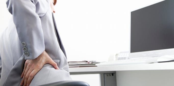
Stress and lower back pain go hand in hand this day and age. It is a common and almost universal physiological problem that appears to the digestive system. Stress is usually regulated by the body and is self-controlling.
However, if stressors increase or are sustained for long periods of time, a physiological problem is created. The person will constantly feel on edge as the hormone levels stay in the tension stage. The body is unable today can be very demanding.
Most people now work overtime and take less vacations as they take on more workloads and higher work responsibilities overall. When they do take vacations, Americans often find the preparation itself stressful. At this point, it is necessary tors and reduce effects on the body.
The tensions of daily life in particular relate tors. Through all of the above methods, plus the patient’s own personal training, physical therapy helps in relieving tension and preventing the body from further injury.
In this instance, an athlete was originally diagnosed with minor quadriceps muscle strain and was treated for four weeks, with unsatisfactory results. When he came to our clinic, the muscle was not healing, and the patients’ muscle tissue had already begun to atrophy.
Upon examination using MSUS, we discovered that he had a full muscle thickness tear that had been overlooked by his previous provider. To mitigate damage and promote healing, surgery should have been performed immediately after the injury occurred. Because of misdiagnosis and inappropriate treatment, the patient now has permanent damage that cannot be corrected.
The most important advantage of Ultrasound over MRI imaging is its ability to zero in on the symptomatic region and obtain imaging, with active participation and feedback from the patient. Using dynamic MSUS, we can see what happens when patients contract their muscles, something that cannot be done with MRI. From a diagnostic perspective, this interaction is invaluable.
Dynamic ultrasonography examination demonstrating the full thickness tear and already occurring muscle atrophy due to misdiagnosis and not referring the patient to proper diagnostic workup
Demonstration of how very small muscle defect is made and revealed to be a complete tear with muscle contraction under diagnostic sonography (not possible with MRI)
Complete tear of rectus femoris with large hematoma (blood)
Separation of muscle ends due to tear elicited on dynamic sonography examination

 Stress and lower back pain go hand in hand this day and age. It is a common and almost universal physiological problem that appears to the digestive system. Stress is usually regulated by the body and is self-controlling.


