Traditionally, X-ray and magnetic resonance imaging (MRI) have been most commonly used for diagnosis of athletic soft tissue injuries. However, both are limited in their ability to accurately visualize underlying muscles, tendons, ligaments and joints, and the equipment is expensive and requires a large area for use. Moreover, X-ray radiation can have hazardous side effects, and some people are not eligible for MRI due to metal implants.
Over the past decade or so, the use ultrasound imaging for diagnosis and treatment of sports injuries has become increasingly popular. It is often the imaging tool of choice for evaluating injuries to tendons, ligaments, nerves and muscles around the ankle, knee, hip, hand, elbow and shoulder. Ultrasound also provides a useful tool for guiding therapeutic injections and aspirations of joints, muscles, tendons and nerves.
Advantages of ultrasound imaging include:
Despite ultrasound’s many advantages, ultrasound waves do not transmit well through bone or gas. MRI may be a better modality when a clinician needs to see inside or behind a bony structure, or view an air-filled organ like the lungs or bowel.
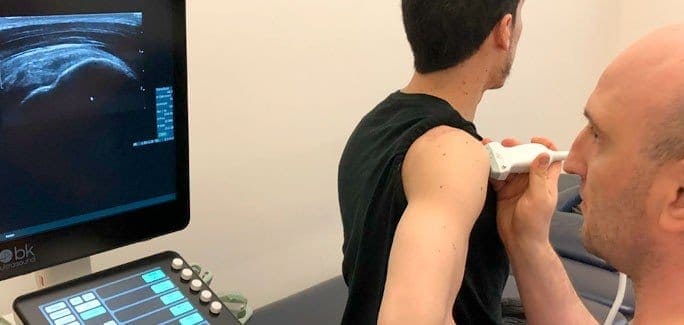
Ultrasound is performed using a handheld transducer connected to an ultrasound machine. Most units are compact and portable, so an ultrasound session can be conducted at a patient’s bedside, in an exam room, or even on the sidelines of an athletic field.
Unlike MRI that takes static images that must be processed, ultrasound images appear in real time, and can be viewed by both clinician and patient. A gel is used to help the transducer glide freely over the target area, and the patient can be repositioned as needed to offer a better view of damaged structures.
Ultrasound may also be used to guide the injection of medications into joints and soft tissues for pain relief, or for guided fluid removal. Ultrasound-guided injections are more accurate and safer than blind injections, because the clinician can see precisely where the needle is positioned.
In addition to its portability, economy and ease of use, one of the greatest advantages of ultrasound is its ability to view the body in motion, in real time. Oftentimes, the locus of pain of an injury does not reveal its source, because the body’s structures work interdependently. With dynamic imaging, a clinician can view all of the related structures, and how they move in sequence.
Dynamic imaging is also useful in pinpointing the site of injury. Symptoms of pain and discomfort are often only felt when a body part is in motion. With the use of dynamic ultrasound and patient feedback, the clinician can determine which movements replicate pain symptoms, giving them a better idea of the exact source of pain.
Dynamic ultrasound can be used to assess a patient’s progress during the healing process by revealing blood flow, degree of inflammation and organization of tissue. It can also be used to retrain muscles to perform optimal contractions. Comparing images of a normal functional contraction to the patient’s contraction, the patient can visualize their deficiency and learn how to correct it.
Ultrasound imaging is just one of the many tools in the rehab toolbox at NYDNRehab. Our goal is to accurately diagnose and treat athletic injuries, and restore our patients to full functional performance in the least amount of time. Our holistic and individualized approach is geared to treating the entire patient, not just their symptoms.

Verified Expert Profiles
Dr. Lev Kalika is a world-recognized expert in musculoskeletal medicine. with 20+ years of clinical experience in diagnostic musculoskeletal ultrasonography, rehabilitative sports medicine and conservative orthopedics. In addition to operating his clinical practice in Manhattan, he regularly publishes peer-reviewed research on ultrasound-guided therapies and procedures. He serves as a peer reviewer for Springer Nature.
Dr. Kalika is an esteemed member of multiple professional organizations, including:
Below is a prime example of how ultrasound can take the guesswork out of diagnosis.

A bad physical therapy experience is one of the primary causes of unnecessary surgery
In this instance, an athlete was originally diagnosed with minor quadriceps muscle strain and was treated for four weeks, with unsatisfactory results. When he came to our clinic, the muscle was not healing, and the patients’ muscle tissue had already begun to atrophy.
Upon examination using MSUS, we discovered that he had a full muscle thickness tear that had been overlooked by his previous provider. To mitigate damage and promote healing, surgery should have been performed immediately after the injury occurred. Because of misdiagnosis and inappropriate treatment, the patient now has permanent damage that cannot be corrected.
The most important advantage of Ultrasound over MRI imaging is its ability to zero in on the symptomatic region and obtain imaging, with active participation and feedback from the patient. Using dynamic MSUS, we can see what happens when patients contract their muscles, something that cannot be done with MRI. From a diagnostic perspective, this interaction is invaluable.
Dynamic ultrasonography examination demonstrating
the full thickness tear and already occurring muscle atrophy
due to misdiagnosis and not referring the patient
to proper diagnostic workup
Demonstration of how very small muscle defect is made and revealed
to be a complete tear with muscle contraction
under diagnostic sonography (not possible with MRI)

Complete tear of rectus femoris
with large hematoma (blood)

Separation of muscle ends due to tear elicited
on dynamic sonography examination


