HomeBlogThe Case Against Valsalva Pushing in the Second Stage of Labor
The Case Against Valsalva Pushing in the Second Stage of Labor
Natural childbirth is as old as time itself…if it weren’t, none of us would be here! For most of human history, childbirth has been the purview of women, mostly midwives and female relatives, who assisted a laboring mother as she did what comes naturally.
However, shortly after World War II, that all changed as modern medical intervention hijacked childbirth and turned it in to a medical condition, subverting the natural biological process and placing it in the hands of mostly male physicians.
As more and more women began giving birth in hospitals, much was lost in terms of spontaneity during labor, and expectant mothers became patients, with all the implications that medical meddling brings with it. In effect, women gave up their rights to the whims and commands of medical personnel.
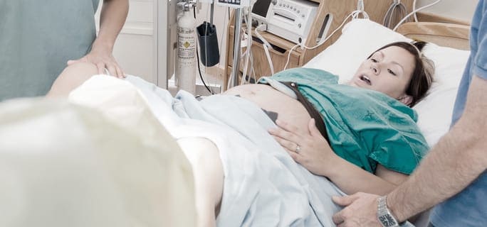
Second-Stage Labor Pushing and Perineal Tearing
One unfortunate and unnecessary outcome of medical childbirth is perineal tearing, often a consequence of second-stage labor pushing, where the mother is encouraged to the baby.
With forced pushing, tearing to take its course.
One study by Kelly et a. (2010) found that delaying pushing for 90 minutes after full cervical dilation actually shortened the time of labor significantly, by a margin of 51%, with no significant increase in overall time for the second stage of labor.
Changing the Way Mothers Labor
One reason perineal tearing became common during medical childbirth is that for decades women were confined to the mother.
Now, new guidelines by the by the Royal College of Obstetricians and Gynaecologists (RCOG) and the Royal College of Midwives in the UK discourage pushing during the second stage of labor and encourage alternative postures, along with breathing through contractions and letting the body and gravity help move the baby through the birth canal. The guidelines also discourage pulling the baby by the shoulders as it can put more pressure on the mother’s perineum.
Physical Therapy for Pre- and Post-Partum Care
Women in peak physical condition are likely to recover more quickly. Working with a physical therapist can help you get your body ready for a safe and beautiful childbirth experience by stabilizing your pelvis and strengthening the muscles of your pelvic floor.
The women’s health professionals at NYDNRehab can assist you in obtaining optimal health in preparation for childbirth, and after the baby is born. Contact NYDNRehab today for a pre-natal or post-partum consultation, and discover the difference physical therapy can make for both you and your baby.
In this instance, an athlete was originally diagnosed with minor quadriceps muscle strain and was treated for four weeks, with unsatisfactory results. When he came to our clinic, the muscle was not healing, and the patients’ muscle tissue had already begun to atrophy.
Upon examination using MSUS, we discovered that he had a full muscle thickness tear that had been overlooked by his previous provider. To mitigate damage and promote healing, surgery should have been performed immediately after the injury occurred. Because of misdiagnosis and inappropriate treatment, the patient now has permanent damage that cannot be corrected.
The most important advantage of Ultrasound over MRI imaging is its ability to zero in on the symptomatic region and obtain imaging, with active participation and feedback from the patient. Using dynamic MSUS, we can see what happens when patients contract their muscles, something that cannot be done with MRI. From a diagnostic perspective, this interaction is invaluable.
Dynamic ultrasonography examination demonstrating the full thickness tear and already occurring muscle atrophy due to misdiagnosis and not referring the patient to proper diagnostic workup
Demonstration of how very small muscle defect is made and revealed to be a complete tear with muscle contraction under diagnostic sonography (not possible with MRI)
Complete tear of rectus femoris with large hematoma (blood)
Separation of muscle ends due to tear elicited on dynamic sonography examination