HomeBlogThe Case for Sonography in Evaluating Injury
The Case for Sonography in Evaluating Injury
Since its advent in the 1980s, Magnetic Resonance Imaging, or MRI, has been broadly used in medicine for the diagnosis of musculoskeletal conditions. Meanwhile, sonography, while emerging and evolving simultaneously, has taken a back seat tool for providing state-of-the art imaging for patients.
MRI vs Ultrasound:
While both MRI and Ultrasound produce an image of the body’s inner landscape, they have several differences:
● Ultrasound uses reflected sound waves to create an image, whereas MRI uses magnetic fields. ● Ultrasound equipment is relatively inexpensive and takes up little space, while MRI is very expensive and requires a large space to house equipment. ● MRI takes a still picture, while Ultrasound gives you a real-time moving image. ● MRI can “see” through bone and air, while ultrasound cannot.
10 Reasons Why Sonography is a Superior Choice for Evaluating Injury
Dr. Nazarian outlines a number of advantages of ultrasound over MRI:
1. Not all patients can safely undergo MRI: Patients with implanted devices like pacemakers and other metal orthopedic implants are not good candidates for MRI, nor are claustrophobic patients, or those who find it difficult or painful to lie supine during MRI. 2. Ultrasound images have higher resolution, revealing more minute detail than MRI, and can detect foreign bodies and calcifications not visible on MRI. 3. Sonography allows for dynamic imaging: Many injuries or musculoskeletal abnormalities only manifest during movement. For example, snapping hip syndrome is evident during hip movement. Dynamic imaging allows the therapist to observe the anomaly in action, something that cannot be done with a static image while the patient lies supine. 4. The ultrasound device can be targeted to a specific area of pain, increasing the chances of detecting the problem and reporting clinically relevant findings, whereas the MRI image is broad and general, increasing the likelihood of misdiagnosis or missed diagnosis. 5. Ultrasound is safe for patients with pacemakers and other implanted metal devices, and it can reveal impingement of soft tissue between orthopedic hardware. 6. Color Doppler sonography and power Doppler sonography offer high resolution imaging that can distinguish between veins and arteries, detect neuromas, identify foreign bodies and depict inflammation and infection, none of which can be seen with MRI. 7. Ultrasound can distinguish between solid material and fluid, reducing the risk of misdiagnosis of cysts or other collections of fluids. 8. Sonography is effective in guiding treatment interventions, targeting soft tissue masses during needle aspiration or core biopsies, and during treatments like injections of corticosteroids, sparing the patient misdirected needle jabs. 9. Sonography makes it easy to the other, giving the clinician a frame of reference during evaluation. 10. The ultrasound probe can be easily moved about the patient’s body, making it a superior choice for evaluating long structures in the body, such as peripheral nerves.
We should also note that MRI imaging is typically conducted by a lab technician, and later viewed by a radiologist without the patient present. Ultrasound evaluation, on the other hand, is very interactive between patient and practitioner, placing the patient at the center of diagnosis, where they are able to provide verbal feedback about pain and discomfort associated with their area of injury.
Since its advent in the 1980s, Magnetic Resonance Imaging, or MRI, has been broadly used in medicine for the diagnosis of musculoskeletal conditions. Meanwhile, sonography, while emerging and evolving simultaneously, has taken a back seat tool for providing state-of-the art imaging for patients. MRI vs Ultrasound: While both MRI and Ultrasound produce an image of the body’s inner landscape, they have several differences:
● Ultrasound uses reflected sound waves to create an image, whereas MRI uses magnetic fields.
● Ultrasound equipment is relatively inexpensive and takes up little space, while MRI is very expensive and requires a large space to house equipment.
● MRI takes a still picture, while Ultrasound gives you a real-time moving image.
● MRI can “see” through bone and air, while ultrasound cannot.
In this instance, an athlete was originally diagnosed with minor quadriceps muscle strain and was treated for four weeks, with unsatisfactory results. When he came to our clinic, the muscle was not healing, and the patients’ muscle tissue had already begun to atrophy.
Upon examination using MSUS, we discovered that he had a full muscle thickness tear that had been overlooked by his previous provider. To mitigate damage and promote healing, surgery should have been performed immediately after the injury occurred. Because of misdiagnosis and inappropriate treatment, the patient now has permanent damage that cannot be corrected.
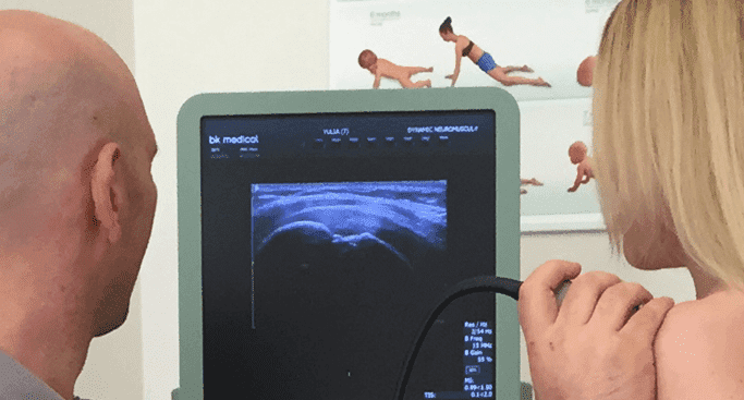
The most important advantage of Ultrasound over MRI imaging is its ability to zero in on the symptomatic region and obtain imaging, with active participation and feedback from the patient. Using dynamic MSUS, we can see what happens when patients contract their muscles, something that cannot be done with MRI. From a diagnostic perspective, this interaction is invaluable.
Dynamic ultrasonography examination demonstrating the full thickness tear and already occurring muscle atrophy due to misdiagnosis and not referring the patient to proper diagnostic workup
Demonstration of how very small muscle defect is made and revealed to be a complete tear with muscle contraction under diagnostic sonography (not possible with MRI)
Complete tear of rectus femoris with large hematoma (blood)
Separation of muscle ends due to tear elicited on dynamic sonography examination