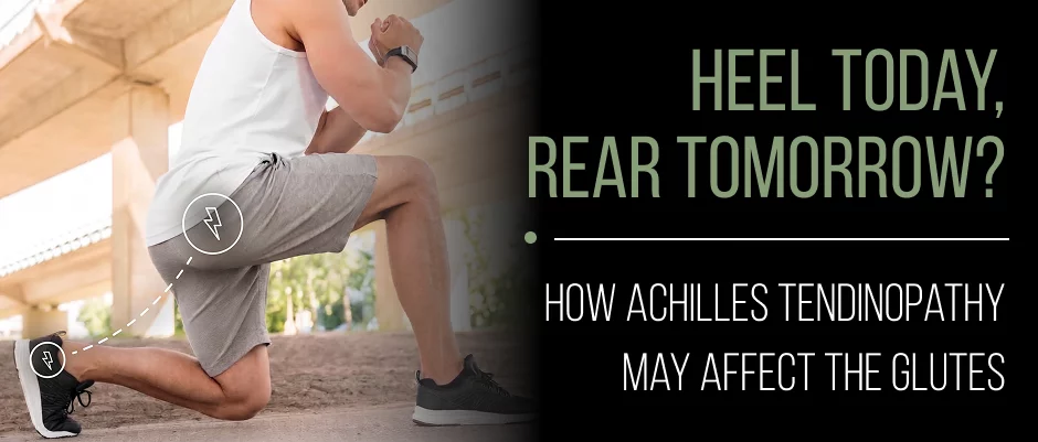
If your heel hurts, the instinct is to treat the heel. But a growing body of sports-medicine research suggests Achilles tendinopathy rarely acts alone, and that what happens at your hips, especially your glutes, may be part of the story.

Not all Achilles pain is the same. Follow this quick guide to help determine the need for professional evaluation.
If you have any of the following symptoms, please seek immediate first aid:
These may be symptoms of a complete rupture of the Achilles tendon which is a surgical emergency requiring immediate evaluation.
If you notice any of these, please make an appointment within 1-2 weeks:
Monitor and self-manage if:
If in any doubt, early assessment will avoid minor overload from developing into chronic tendinopathy.
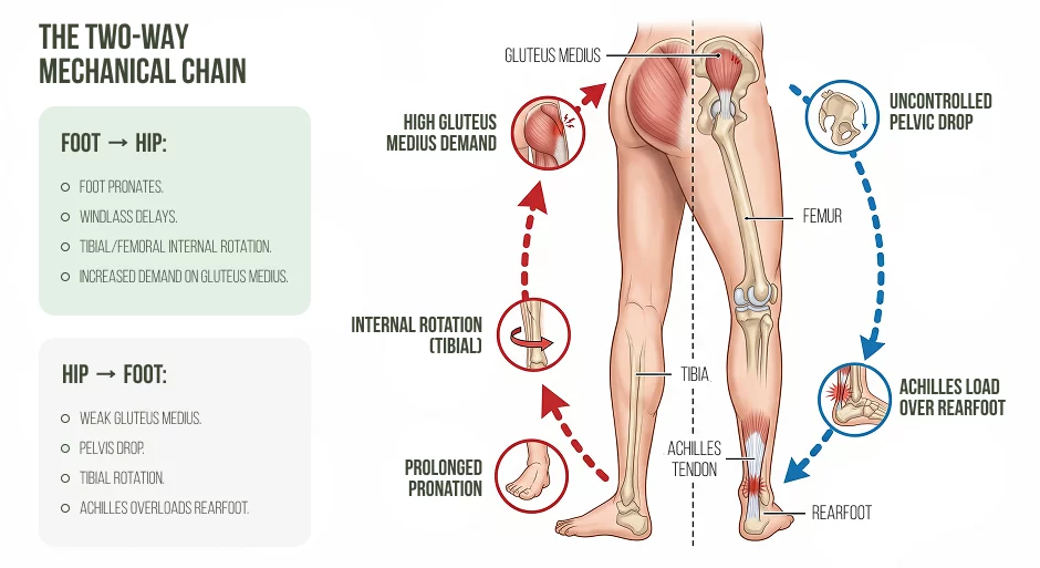
It is important to be familiar with one of the basic rules before starting to do research. The Achilles tendon is not a standalone tendon. It is located in the middle of a mechanical chain that connects the toes to the foot, ankle, knee, hip and pelvis. This chain is used for both the up and down forces, moving up from the foot and down from the hip, on each and every stride.
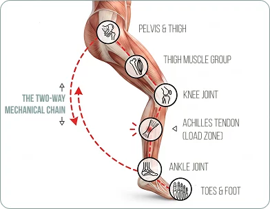
This is known as the Two-Way Chain. If one of the links in this chain is weak, stiff or poorly timed, the load is not just lost. It is redirected. One of the most frequent locations for that redirected load to build up until the tissue’s capacity is exceeded is the Achilles tendon.
If you are injured, your first thought is to think about the part of your body that hurts. A runner with Achilles pain ices his heel, stretches his calf and hopes for the best. However, physicians and scientists are gaining more knowledge that injuries do not occur in isolation.
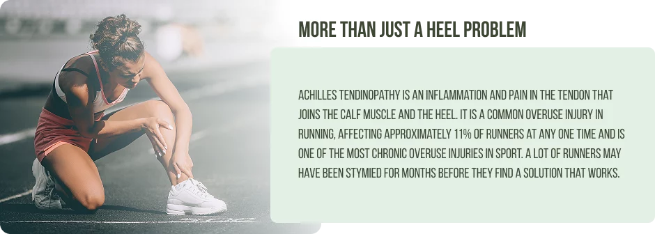
Achilles tendinopathy is an inflammation and pain in the tendon that joins the calf muscle and the heel. It is a common overuse injury in running, affecting approximately 11% of runners at any one time and is one of the most chronic overuse injuries in sport. A lot of runners may have been stymied for months before they find a solution that works.
Modern sports medicine recognizes the body as a kinetic chain. Cumulative overload has often occurred when it has passed a tissue’s biological capacity and the tissue is painful, but not necessarily the cause of the dysfunction.
The Achilles tendon is the biggest and strongest tendon in the human body. It acts like a biological spring, storing and releasing elastic energy during running, and absorbing forces 6–8 times greater than body weight per stride.
When tendons are loaded, they remodel their collagen structure in order to become healthier. As training slowly progresses, the tendon gets stronger. As loading exceeds the tendon’s ability to adapt, the process breaks down and results in disorganized collagen, abnormal tissue changes and chronic pain.
Common contributors include:





Treating the tendon alone will not cure the underlying problem.
Perhaps Achilles tendinopathy can have repercussions that ascend up the kinetic chain, all the way to the buttocks.
Take a look at the findings of a study published in October 2013 by Medicine & Science in Sports & Exercise, published by the American College of Sports Medicine. This study included 33 male runners, 19 healthy and 14 with Achilles tendinopathy.
The researchers observed the following:
This is a small sample size, an important limitation as there were only 14 participants in the tendinopathy group. The results presented should be regarded as hypothesis-generating, not as conclusions. They see a pattern that has some significance, but not a universal pattern.
The study found that there was a correlation between Achilles tendinopathy and delayed activation of the gluteals. It did not prove that one caused the other. What it shows is that runners with Achilles tendinopathy have some differences in their running gait, and that these differences are associated with measurable differences in gluteal muscle recruitment.
The clinical effect of the delay, whether it be due to the nature of the drug or the patient’s condition, is the same. When the gluteus medius fires late it is not going to be effective at controlling pelvic drop. A late firing gluteus maximus will not function optimally during hip extension and propulsion. Both gaps impose a larger force on the calf and Achilles tendon to deal with forces that should have been dealt with higher up the chain.
 Are weak or delayed glutes a cause of Achilles tendinopathy?Or do weak glutes cause Achilles tendinopathy?Which came first?Is there any muscle other than the glutes that can cause Achilles pain?
Gluteus Maximus
The gluteus maximus is the largest muscle in the human body. It is responsible for hip extension during running, for generating push-off force, for deceleration of the limb at initial contact and for stabilizing the pelvis. It attaches to the thoracolumbar fascia, which is a vital force transmission pathway between the pelvis and the posterior oblique sling, and is involved in the transmission of forces with each running stride.
With impaired function of gluteus maximus, propulsive demand moves distally to the calf and Achilles tendon. This redistribution over thousands of strides is a significant increase in cumulative tendon stress.
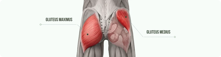
Gluteus Medius
During single-leg stance (about 40% of running cycle), the gluteus medius stabilizes the pelvis. If it fires late or doesn’t shoot enough, the pelvis falls to the unsupported side.
This sets in a mechanical chain; the femur adducts and internally rotates, the tibia follows and the back foot is pushed into over-pronation. This foot position will alter the angle of the Achilles tendon loading, resulting in greater stress over time.
It should be noted that changes in the mechanics of the frontal plane do not necessarily lead to tendinopathy. There are many runners who have some weakness in their gluteus medius but don’t experience any symptoms. The relationship is probabilistic not deterministic.
The foot must be able to absorb impact forces when loading and change to a rigid propulsive lever in a fraction of a second during push-off.
This is achieved by the windlass mechanism. When the heel rises and the toes dorsiflex, the plantar fascia tightens, raising the medial arch and making the foot stiffer for push-off. This mechanism is disrupted if there is a limitation in hallux dorsiflexion, such as hallux rigidus or hallux limitus. The Achilles tendon will then have to produce more plantarflexion force to compensate.
The plantar fascia and Achilles tendon both attach to the calcaneus. Cadaveric studies have shown that these structures are continuous with each other via fascia. One affects the other and coexisting plantar fascia pathology is clinically important in runners with Achilles tendinopathy.
The Two-Way Chain at the foot is used as follows:
Both directions need to be evaluated and resolved.
The foot must be able to absorb impact forces when loading and change to a rigid propulsive lever in a fraction of a second during push-off.
This is achieved by the windlass mechanism. When the heel rises and the toes dorsiflex, the plantar fascia tightens, raising the medial arch and making the foot stiffer for push-off. This mechanism is disrupted if there is a limitation in hallux dorsiflexion, such as hallux rigidus or hallux limitus. The Achilles tendon will then have to produce more plantarflexion force to compensate.
The plantar fascia and Achilles tendon both attach to the calcaneus. Cadaveric studies have shown that these structures are continuous with each other via fascia. One affects the other and coexisting plantar fascia pathology is clinically important in runners with Achilles tendinopathy.
Rehabilitation Principles
Successful rehabilitation restores tendon capacity and corrects the mechanical environment that put it under stress. Both are not enough.
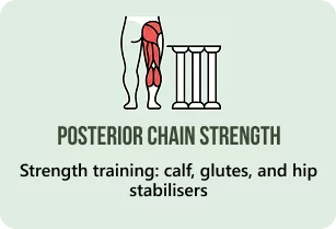
Progressive tendon loading is the cornerstone. Current evidence supports:

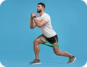
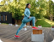
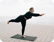
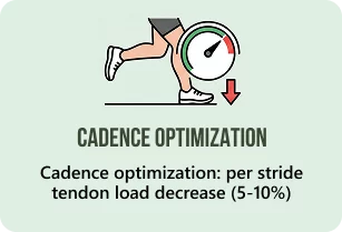
Gait retraining corrects the mechanical contributors that were identified in assessment. Modifying running cadence to increase it by 5–10% decreases peak Achilles tendon loading per stride and is a simple, inexpensive modification for many runners.
Stretching calves will NOT restore tissue quality to the tendon, correct any biomechanical drivers, or restore load-bearing capacity. In insertional tendinopathy, excessive stretching may create symptoms by exacerbating compressive forces at the calcaneal insertion. Stretching is used to support other treatments, but is not a main treatment.
A lack of pain inhibition during effective muscle recruitment, a mechanical barrier because of structural pathology (partial gluteus medius tear, labral lesion, hallux rigidus), or training load that is too high for the tendon to net biological recovery are all potential reasons for failure of strengthening programs. If progress is not being made, then re-evaluate to determine if these barriers are present.
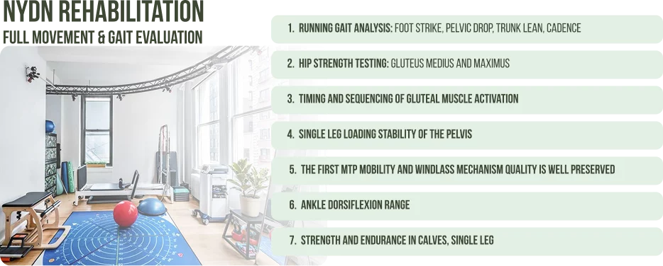
Two people with the same symptoms can have very different underlying causes and very different treatment options.
If further diagnosis is required, diagnostic ultrasound is our preferred imaging modality of the first choice as it allows the identification of tendon thickening, structural disorganisation, neovascularisation and partial tears in real time. MRI is only used for complex presentations or surgical planning.
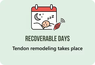
Tendon remodeling is a slow process. The collagen turnover in tendon is measured in months, which is much slower than the response of muscle to training within weeks. Early care for runners leads to faster and fuller recovery. The most frequent cause of setback is returning to full training too early.
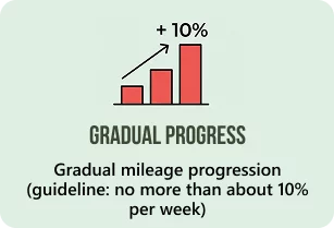
Practical prevention strategies:
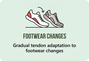
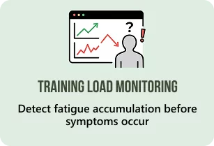
In either case, the doctor and physical therapist should always take the function of the gluteal muscles into account when evaluating and treating Achilles tendon issues. The 2013 study is part of a series of studies that has been ongoing for some time that running injuries are not usually singular occurrences.
The evidence is sufficient to act upon: The Achilles tendon is part of a system that starts from the trunk and goes to the toes. The first step in successful treatment is to understand each runner’s dysfunctional pattern, rather than treating the painful tendon itself.
Our sports medicine and physical therapy team at NYDN Rehabilitation treats Achilles tendinopathy at its root by employing a thorough movement assessment program, advanced diagnostic ultrasound and personalized rehabilitative strategies. If you are a runner who has heel or tendon pain that persists, give us a call for an appointment.
Request an appointment»

Our location: 11 West 25th Street 5th floor, New York, NY 10010
Dr. Lev Kalika is a world-recognized expert in musculoskeletal medicine. with 20+ years of clinical experience in diagnostic musculoskeletal ultrasonography, rehabilitative sports medicine and conservative orthopedics. In addition to operating his clinical practice in Manhattan, he regularly publishes peer-reviewed research on ultrasound-guided therapies and procedures. He serves as a peer reviewer for Springer Nature.
Dr. Kalika is an esteemed member of multiple professional organizations, including:

Below is a prime example of how ultrasound can take the guesswork out of diagnosis.
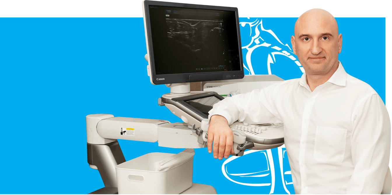

A bad physical therapy experience is one of the primary causes of unnecessary surgery

In this instance, an athlete was originally diagnosed with minor quadriceps muscle strain and was treated for four weeks, with unsatisfactory results. When he came to our clinic, the muscle was not healing, and the patients’ muscle tissue had already begun to atrophy.
Upon examination using MSUS, we discovered that he had a full muscle thickness tear that had been overlooked by his previous provider. To mitigate damage and promote healing, surgery should have been performed immediately after the injury occurred. Because of misdiagnosis and inappropriate treatment, the patient now has permanent damage that cannot be corrected.
The most important advantage of Ultrasound over MRI imaging is its ability to zero in on the symptomatic region and obtain imaging, with active participation and feedback from the patient. Using dynamic MSUS, we can see what happens when patients contract their muscles, something that cannot be done with MRI. From a diagnostic perspective, this interaction is invaluable.
Dynamic ultrasonography examination demonstrating
the full thickness tear and already occurring muscle atrophy
due to misdiagnosis and not referring the patient
to proper diagnostic workup
Demonstration of how very small muscle defect is made and revealed
to be a complete tear with muscle contraction
under diagnostic sonography (not possible with MRI)
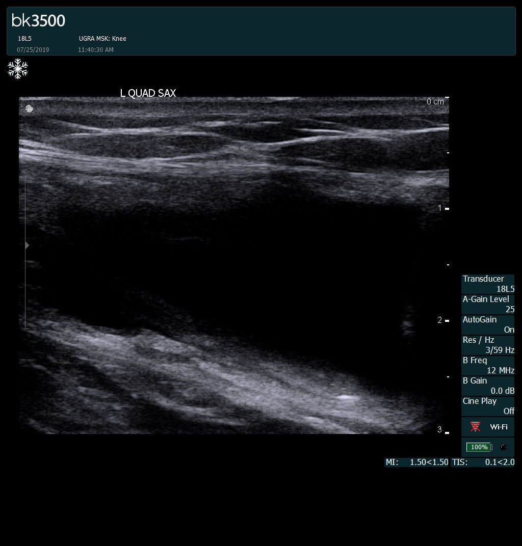
Complete tear of rectus femoris
with large hematoma (blood)
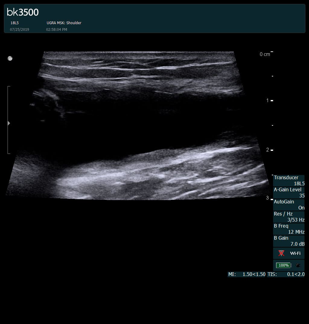
Separation of muscle ends due to tear elicited
on dynamic sonography examination

 Request Telehealth