Hamstring Injury

benefits_of_shocking_vawe_1

Hamstring injuries are among the most common injuries suffered by athletes, especially in sports like soccer and American football that require rapid acceleration, sudden stops and quick directional changes. Sports fans cringe to see an athlete crumple to the field, clutching a hamstring in obvious pain. The pain is sometimes so severe that the athlete will need to be carried off the field.

Anatomy of the Hamstring Muscles

The hamstrings are made up of three muscles in the back of the thigh. They include the short and long heads of the biceps femoris, the semitendinosus and the semimembranosus. The short and long heads of the biceps femoris originate from closely placed sites on the posterior/inferior surface of the ischial tuberosity. They both converge to insert onto the fibular head, while the semimembranosus and the semitendinosus insert on the proximal medial tibia.

benefits_of_shocking_vawe_1

The hamstring muscles cross over both the hip and knee joints, contributing to both knee flexion and hip extension. The hamstrings stabilize the hip joint during movement, and act together with the collateral ligaments to stabilize the knee joint.

Symptoms

Hamstring injuries can range from a minor bruise or strain to a full scale rupture.

The athlete may have difficulty walking and standing, and exercise may be painful or impossible. Popping sounds during movement are common after a hamstring injury. If there is a serious rupture movement involving the lower extremities may be impossible, and the injured leg will not be able to bear the patient’s weight.

Diagnosis

app

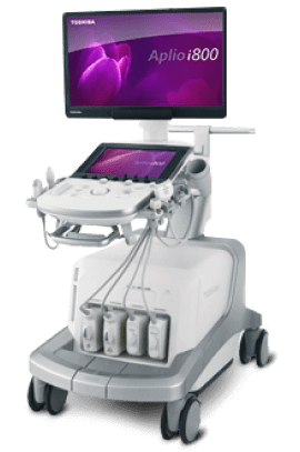
Hamstring muscle injuries must be radiologically examined with the help of MRI and ultrasound to classify them into proper categories, based on their anatomical location. For hamstring muscle injuries, MRI is more comprehensive than ultrasound, but ultrasound is better in the initial stages, and useful for removal of an effusion using ultrasound guidance. Ultrasound is also better for reexamination and gauging the healing process, because it can be performed multiple times in a matter of minutes. It is up to your physician to decide which modality to perform and when.

Explore more advanced diagnostic tools available only at NYDNRehab:

img

Grading Hamstring Muscle Injuries

In the past, hamstring muscle injuries were graded by severity using a very simple system. More recently, due to improvements in ultrasound and MRI, hamstring muscle injuries can be classified based on bone-tendo, tendon-fascia, tendon-muscle and muscle-fascia interactions.This anatomical division allows for better treatment and a more precise estimation of when an athlete can return to the field.

Treatment

A ruptured hamstring requires immediate medical attention. Mild injuries can be treated with the PRICE procedure (protect, rest, ice, compress and elevate), but the injured patient should still consult a sports physician or physical therapist.

For a mild injury, the doctor will likely prescribe analgesics and refer the patient to a physical therapist. If the hamstring is ruptured, surgery may be needed to suture the damaged tissue together. After that, the patient will need to wear a brace and rest for about six weeks or until the hamstring is ready for rehab.

In preparation for physical therapy exercises, the therapist may provide therapeutic massage, or wrap the area to protect the muscle from overuse. Rehab may include graded eccentric loading exercises, along with extrinsic feedback exercises to restore brain-muscle connectivity.

Sometimes, it is impossible to make a trip to the clinic. Whether you’re confined at home or on the go, NYDNRehab TeleHealth services provide a convenient way to receive ongoing professional physical therapy and chiropractic treatment at home, in the office or while traveling. With TeleHealth, you never need to miss a physical therapy session or lose hard-earned gains in treatment.

If you intend to maintain a physically active lifestyle, the sports medicine pros at NYDNRehab can help you rehabilitate your injury so you can return to play. Our unique high-tech gait lab is designed to identify and quantify motor deficits and correct them before injuries occur. Contact us today, and achieve your peak performance potential while reducing your risk of injury. At NYDNRehab, movement is life!

About the Author

Dr. Lev Kalika is clinical director of NYDNRehab, located in Manhattan. Lev Kalika is the author of multiple medical publications and research, and an international expert in the field of rehabilitative sonography, ultrasound guided dry needling and sports medicine Dr. Kalika works with athletes, runners, dancers and mainstream clients to relieve pain, rehabilitate injuries, enhance performance and minimize the risk of injuries. His clinic features some of the most technologically advanced equipment in the world, rarely found in a private clinic.

bg

In this instance, an athlete was originally diagnosed with minor quadriceps muscle strain and was treated for four weeks, with unsatisfactory results. When he came to our clinic, the muscle was not healing, and the patients’ muscle tissue had already begun to atrophy.

Upon examination using MSUS, we discovered that he had a full muscle thickness tear that had been overlooked by his previous provider. To mitigate damage and promote healing, surgery should have been performed immediately after the injury occurred. Because of misdiagnosis and inappropriate treatment, the patient now has permanent damage that cannot be corrected.

The most important advantage of Ultrasound over MRI imaging is its ability to zero in on the symptomatic region and obtain imaging, with active participation and feedback from the patient. Using dynamic MSUS, we can see what happens when patients contract their muscles, something that cannot be done with MRI. From a diagnostic perspective, this interaction is invaluable.

Dynamic ultrasonography examination demonstrating
the full thickness tear and already occurring muscle atrophy
due to misdiagnosis and not referring the patient
to proper diagnostic workup

Demonstration of how very small muscle defect is made and revealed
to be a complete tear with muscle contraction
under diagnostic sonography (not possible with MRI)

image

Complete tear of rectus femoris
with large hematoma (blood)

image

Separation of muscle ends due to tear elicited
on dynamic sonography examination

bg
Buy now 3D Gait
Payment Success
Request Telehealth Request Telehealth Request in office visit Book now