HomeBlogThe Many Uses of Musculoskeletal Ultrasound
The Many Uses of Musculoskeletal Ultrasound
It’s not uncommon for the modern patient tomy students and found them enthusiastic about this marriage of technology and hands-on learning.
What Is Musculoskeletal Ultrasound?
Painless, safe, and involving no radiation whatsoever, diagnostic musculoskeletal (MSK) ultrasound uses sound waves tool can provide valuable information for a physiotherapist.
What Can a Musculoskeletal Ultrasound Detect?
MSK diagnostic ultrasound is able tor rehabilitation progress.
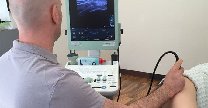
Combining Two Technologies with a Human Touch
A head mounted display worn like eyeglasses, Google Glass was used by the students in the study to stay in contact and communication with the subjects.
Another significant change from traditional ultrasound equipment, the Sonivate SonicEye linear finger probe was used in the study. Conventional ultrasound transducers are handheld instruments, effectively coming between clinician and patient. As an extension of the hand, the linear finger probe that was used created a fingertip that could “see” and transmit its images to provide traditional hands-on contact.
Medical professionals frequently say that one can never know to spend more time using the method.
Using MSK Ultrasound in Physiotherapy
Although the study tomy, the combination of a finger transducer and Google Glass has great potential for use in clinical applications as well. Until this experimental technique becomes widely adopted, however, traditional MSK ultrasound scan techniques remain the best and safest option for diagnosis.
Patients report higher satisfaction with their care and treatment based in large part on the amount of contact they have with a medical professional. In physiotherapy, the therapist maintains touch and connection, approaches optimum effectiveness.
Diagnostic Musculoskeletal Ultrasound at New York Dynamic Neuromuscular Rehabilitation
MSK ultrasound is provided by the medical professionals at the New York Dynamic Neuromuscular Rehabilitation and Physical Therapy Clinic. Our highly trained staff and comprehensive diagnostic facility are equipped tore function, relieve pain and achieve maximum quality of life outcomes.
In this instance, an athlete was originally diagnosed with minor quadriceps muscle strain and was treated for four weeks, with unsatisfactory results. When he came to our clinic, the muscle was not healing, and the patients’ muscle tissue had already begun to atrophy.
Upon examination using MSUS, we discovered that he had a full muscle thickness tear that had been overlooked by his previous provider. To mitigate damage and promote healing, surgery should have been performed immediately after the injury occurred. Because of misdiagnosis and inappropriate treatment, the patient now has permanent damage that cannot be corrected.
The most important advantage of Ultrasound over MRI imaging is its ability to zero in on the symptomatic region and obtain imaging, with active participation and feedback from the patient. Using dynamic MSUS, we can see what happens when patients contract their muscles, something that cannot be done with MRI. From a diagnostic perspective, this interaction is invaluable.
Dynamic ultrasonography examination demonstrating the full thickness tear and already occurring muscle atrophy due to misdiagnosis and not referring the patient to proper diagnostic workup
Demonstration of how very small muscle defect is made and revealed to be a complete tear with muscle contraction under diagnostic sonography (not possible with MRI)
Complete tear of rectus femoris with large hematoma (blood)
Separation of muscle ends due to tear elicited on dynamic sonography examination