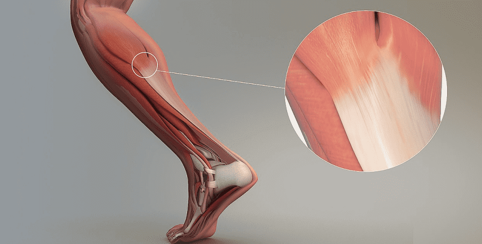
Tendons are tough connective tissues that are continuous with muscles, attaching them securely to your bones. When a muscle shortens during contraction, it pulls on its tendon which in turn pulls on its associated bone, producing movement. When subjected to high force loads during exercise, sports or everyday activities, tendons can become injured and inflamed, causing pain and impeding movement.
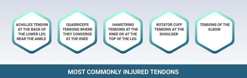
Although tendons and their associated muscles are found throughout the human body, some are more vulnerable to injury than others. Most tendon injuries occur near joints, including the ankle, knee, hip, shoulder and elbow. While acute tendon injuries do occur, most develop over time as repetitive overuse injuries. Tendons may also become injured due to a fall or a sudden unintentional movement.
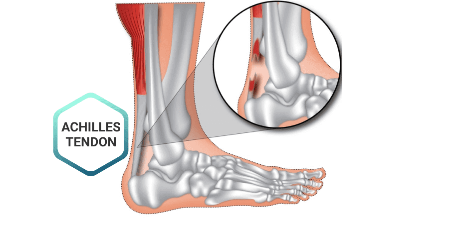
Some of the most commonly injured tendons include:
Due to a lower blood supply and ongoing use, tendon tissue can be slow to heal. Physical therapy offers a number of treatment approaches to accelerate tendon healing and restore normal movement.
The primary symptom of tendon injury is pain, swelling and tenderness near the joint. Other symptoms include:
Diagnosis of tendon injuries begins with a health history and physical exam. Your therapist will ask you questions about your physical activities, lifestyle, when your pain began, and other relevant questions. They will palpate the tendon area and do some functional tests for strength and range of motion.
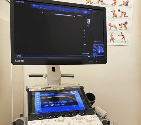
Diagnostic ultrasonography is the most effective imaging method for viewing tendons and their surrounding structures. It enables the clinician to view the tendon in real time, with the patient in motion. Unlike static imaging technologies like Xray and MRI, musculoskeletal ultrasound allows for scanning the entire length of a tendon with the joint in motion, while the patient provides verbal feedback. Because results are instantaneous, treatment can begin immediately.
Despite tendon injuries being fairly common, there is much controversy about the best way to rehabilitate them. While much theoretical research has been conducted, there are very few high-quality clinical trials that show significant improvement from specific treatments.
The main goal of treatment is to improve the ability of the muscle and tendon to manage load. Because muscles and tendons work as a musculotendinous unit, the tendon cannot be treated independently of the muscle. Muscle loading during rehab should consider the nature, speed and magnitude of forces necessary to restore full funcion, particularly when an athlete intends to return to play.
A variety of loading approaches have been used with varying degrees of success. Eccentric loading is fairly popular, where loading is accentuated during the muscle lengthening phase. Isometrics have also been used in combination with concentric and eccentric loading protocols. In isometric exercise, muscle tension is present, but there is no change in joint angle or muscle length.
One study published by the British Journal of Sports Medicine found that isometric exercise reduced pain and promoted healing of the patellar tendon, while eccentric exercises were painful and ineffective. However, it is likely that the effectiveness of different loading methods varies with the tendon location, injury type and the individual patient.
The sports medicine specialists at NYDNRehab are dedicated to eliminating tendon pain and speeding up the healing process. Whether you are a competitive athlete wanting to return to sport or a recreational exerciser who wants to stay active, treating your tendon pain is paramount.
Our integrative and comprehensive examination includes:
Our treatment options include:

Verified Expert Profiles
Dr. Lev Kalika is a world-recognized expert in musculoskeletal medicine. with 20+ years of clinical experience in diagnostic musculoskeletal ultrasonography, rehabilitative sports medicine and conservative orthopedics. In addition to operating his clinical practice in Manhattan, he regularly publishes peer-reviewed research on ultrasound-guided therapies and procedures. He serves as a peer reviewer for Springer Nature.
Dr. Kalika is an esteemed member of multiple professional organizations, including:
Below is a prime example of how ultrasound can take the guesswork out of diagnosis.

A bad physical therapy experience is one of the primary causes of unnecessary surgery
In this instance, an athlete was originally diagnosed with minor quadriceps muscle strain and was treated for four weeks, with unsatisfactory results. When he came to our clinic, the muscle was not healing, and the patients’ muscle tissue had already begun to atrophy.
Upon examination using MSUS, we discovered that he had a full muscle thickness tear that had been overlooked by his previous provider. To mitigate damage and promote healing, surgery should have been performed immediately after the injury occurred. Because of misdiagnosis and inappropriate treatment, the patient now has permanent damage that cannot be corrected.
The most important advantage of Ultrasound over MRI imaging is its ability to zero in on the symptomatic region and obtain imaging, with active participation and feedback from the patient. Using dynamic MSUS, we can see what happens when patients contract their muscles, something that cannot be done with MRI. From a diagnostic perspective, this interaction is invaluable.
Dynamic ultrasonography examination demonstrating
the full thickness tear and already occurring muscle atrophy
due to misdiagnosis and not referring the patient
to proper diagnostic workup
Demonstration of how very small muscle defect is made and revealed
to be a complete tear with muscle contraction
under diagnostic sonography (not possible with MRI)

Complete tear of rectus femoris
with large hematoma (blood)

Separation of muscle ends due to tear elicited
on dynamic sonography examination


