HomeBlogUnderstanding Abdominal Diastasis: What to do, and what to avoid
Understanding Abdominal Diastasis: What to do, and what to avoid
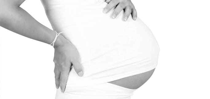
Abdominal diastasis, the separation of the rectus abdominis (six-pack) muscle along the linea alba ligament, is most commonly seen in pregnant and post-partum women. However, diastasis rectus abdominis (DRA) can also occur in people who are overweight and obese, and in people who do strenuous work. It can even occur in fit populations where the abdominal muscles are overworked, or are exercised strenuously with faulty technique.
The linea alba is fibrous, with limited elasticity. During pregnancy, the release of the relaxin hormone enables ligaments throughout the mother’s body to become more elastic and pliable, making room for the baby to grow, and enabling safe passage along the birth canal. Overweight and fit populations who are not pregnant do not have the hormonal advantage of pregnant women, meaning the linea alba is less elastic and more vulnerable to rupture.
Strategies to Avoid DRA
If you want to avoid abdominal diastasis, there are several precautionary measures you can take:
If you are planning to become pregnant, build your core and pelvic floor strength, especially the transverse abdominal muscle.
Avoid excessive abdominal weight gain by exercising, eating whole foods and staying hydrated.
Practice good posture to take pressure off the abdominal canister.
Avoid exercises that increase pressure in your abdomen.
Treating DRA
If you have DRA immediately after pregnancy, it is likely to resolve itself over the course of several weeks. If DRA persists, consider conservative therapy before seeking surgical intervention. In many cases, a physical therapist can work with you to realign the muscles and structures in your abdominal and pelvic region, to reduce DRA and address other common problems associated with it, including incontinence and uterine prolapse.
If you have DRA associated with obesity, you should begin a weight loss program and consult with a physical therapist to determine the best course of treatment.
If your DRA is associated with strenuous workloads or fitness activities, a therapist can help you change the way you work or exercise, and they can recommend strategies for healing and avoiding further damage.
Hypopressive Exercise for DRA
Hypopressive, or low pressure exercises improve the integrity of your abdominal canister without putting excessive pressure on your abdominal wall. When performed correctly and consistently, hypopressive exercises can improve your posture, eliminate incontinence and correct uterine prolapse. They may also be effective in resolving or reducing your DRA.
The sports medicine professionals at NYDNRehab use the most advanced technologies and therapies to diagnose and treat DRA. Once the scope and nature of your DRA have been determined, our physical therapy team will devise an individualized treatment plan for you that will likely include hypopressive exercises, along with other exercises aimed at correcting postural and movement deficiencies.
You do not have to live with DRA. Contact NYDNRehab today to schedule a consultation. You will soon see why NYDNRehab is considered to be the very best rehabilitation clinic in NYC.
In this instance, an athlete was originally diagnosed with minor quadriceps muscle strain and was treated for four weeks, with unsatisfactory results. When he came to our clinic, the muscle was not healing, and the patients’ muscle tissue had already begun to atrophy.
Upon examination using MSUS, we discovered that he had a full muscle thickness tear that had been overlooked by his previous provider. To mitigate damage and promote healing, surgery should have been performed immediately after the injury occurred. Because of misdiagnosis and inappropriate treatment, the patient now has permanent damage that cannot be corrected.
The most important advantage of Ultrasound over MRI imaging is its ability to zero in on the symptomatic region and obtain imaging, with active participation and feedback from the patient. Using dynamic MSUS, we can see what happens when patients contract their muscles, something that cannot be done with MRI. From a diagnostic perspective, this interaction is invaluable.
Dynamic ultrasonography examination demonstrating the full thickness tear and already occurring muscle atrophy due to misdiagnosis and not referring the patient to proper diagnostic workup
Demonstration of how very small muscle defect is made and revealed to be a complete tear with muscle contraction under diagnostic sonography (not possible with MRI)
Complete tear of rectus femoris with large hematoma (blood)
Separation of muscle ends due to tear elicited on dynamic sonography examination