August 14, 2023
A painful condition called coccydynia is truly difficult tolerate. It actually is prompted by inflammation situated in your tailbone. Despite all the guessing about it, the most frequent reason for it is plain trauma a fracture, a heavy fall.
This non-sto coccyx pain interference with your daily life.
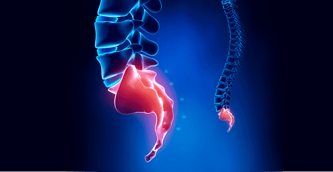
Spinal column ends with coccyx. It has a triangle shape, the bones in it are closely fused.
Traditional medicine considers coccyx to be the remainder of a tail, thus the name for it. EVen though fused tightly, coccyx bones are connected by fibrous tissue.
The allow for slight forward and backward movement that coordinates with movement of the pelvis, hips, and legs. Gluteus maximus muscle attaches to the posterior coccyx’ side, playing an important role in its position.
When sitting or standing, your coccyx, along with every other pelvic basin bone, adjusts position. It literally helps to balance your body and provide balance. This role coccyx plays in human vestibular system is one of the main reasons why removal surgery is so rarely recommended.
In the seated position your body dispenses the overall weight among ischium bones for stability maintenance. The tail bone also acts as a site of attachment for other minor body structures in the area. It is also an insertion point for some of your pelvic floor muscles.
Pain in this condition is generally point-tender, not radiating. In addition towards the front of a chair.
A heavy fall backwards or a complicated delivery of a child can cause Coccydynia. However, about one-third of cases are idiopathic in nature, meaning they arise without a specific identifiable cause. Obesity is also a major factor. Females have an increased risk of developing the condition. It is associated with post childbirth consequences in many cases.
Increase in sympto. Sitting on something really hard may impact and worsen Coccydynia. If the inflammation is long while neglected – having a bowel movement or a sexual intercourse may induce the heavy pain in the area.
Coccydynia is normally diagnosed through medical histo be difficult during the period.
Conservative multifaceted non-invasive treatment for inflammation in tailbone area is common for medical science. Treatments will include the following:
Surgical Remedy for Coccydynia In extreme cases where conservative treatments do not alleviate the extreme pain sensation. Coccygecto a year, and sitting is difficult during the course of healing.
The sports medicine professionals at NYDNRehab have extensive experience treating tailbone pain. We use cutting edge technology, combining diagnostic ultrasonography and extracorporeal shockwave therapy to recovery from coccydynia.

Verified Expert Profiles
Dr. Lev Kalika is a world-recognized expert in musculoskeletal medicine. with 20+ years of clinical experience in diagnostic musculoskeletal ultrasonography, rehabilitative sports medicine and conservative orthopedics. In addition to operating his clinical practice in Manhattan, he regularly publishes peer-reviewed research on ultrasound-guided therapies and procedures. He serves as a peer reviewer for Springer Nature.
Dr. Kalika is an esteemed member of multiple professional organizations, including:
Below is a prime example of how ultrasound can take the guesswork out of diagnosis.

A bad physical therapy experience is one of the primary causes of unnecessary surgery
In this instance, an athlete was originally diagnosed with minor quadriceps muscle strain and was treated for four weeks, with unsatisfactory results. When he came to our clinic, the muscle was not healing, and the patients’ muscle tissue had already begun to atrophy.
Upon examination using MSUS, we discovered that he had a full muscle thickness tear that had been overlooked by his previous provider. To mitigate damage and promote healing, surgery should have been performed immediately after the injury occurred. Because of misdiagnosis and inappropriate treatment, the patient now has permanent damage that cannot be corrected.
The most important advantage of Ultrasound over MRI imaging is its ability to zero in on the symptomatic region and obtain imaging, with active participation and feedback from the patient. Using dynamic MSUS, we can see what happens when patients contract their muscles, something that cannot be done with MRI. From a diagnostic perspective, this interaction is invaluable.
Dynamic ultrasonography examination demonstrating
the full thickness tear and already occurring muscle atrophy
due to misdiagnosis and not referring the patient
to proper diagnostic workup
Demonstration of how very small muscle defect is made and revealed
to be a complete tear with muscle contraction
under diagnostic sonography (not possible with MRI)

Complete tear of rectus femoris
with large hematoma (blood)

Separation of muscle ends due to tear elicited
on dynamic sonography examination


