HomeBlogWeak in the Knees: Osteoarthritis Doubles Among 21st Century Americans
Weak in the Knees: Osteoarthritis Doubles Among 21st Century Americans
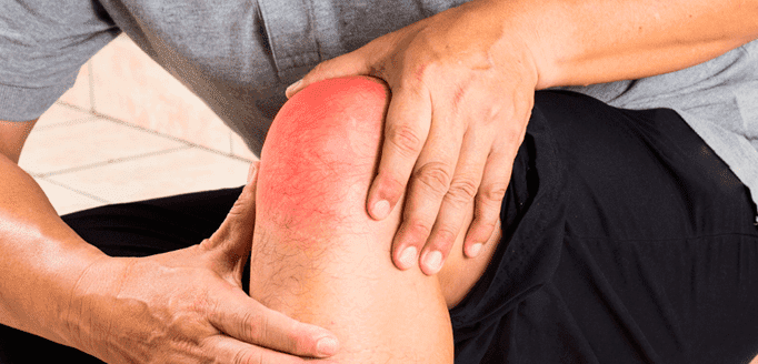
Knee osteoarthritis (OA) is the most common and debilitating joint condition in the United States, affecting more than 19% of adults over age 45. That statistic is not only high, but it is alarming when considering it has more than doubled since the mid-20th Century. Scientists have attempted to the increased incidence of knee OA.
New Study Demolishes Wear and Tear Theory
In 2017, a research team led by Ian J. Wallace wanted to greater stresses placed on the knees over the course of a lifetime.
The research team looked at the knees of cadavers over age 50, scanning prehisto the present, with the oldest knees dating back around 6000 years. OA was diagnosed based on the presence of eburnation, the polishing evident from bone-on-bone contact seen in OA.
The study samples (n = 2,576) included:
Early industrial individuals over age 50 (n = 1,581) who were inhabitants of Cleveland, Ohio and St. Louis, Missouri and who died between 1905 and 1940
Postindustrial individuals (n = 819) who lived in Albuquerque, New Mexico and Knoxville, Tennessee, who died between 1976 and 2015.
Prehisto be over age 50, who were hunter-gatherers (n = 116) and early farmers (n = 60).
Study Findings
After data analysis, the research team found that:
Knee OA was higher among individuals from the postindustrial era compared with individuals from early industrial and prehistoric times
Females were more affected than males
After controlling for sex, postindustrial knee OA prevalence was 16%, 2.6 times higher that the early industrial sample (6%) and 2 times higher than the prehistoric sample (8%)
Among postindustrial individuals with knee OA, 42% had OA in both knees, 2.5 times higher than the prehistoric sample (17%) and 1.4 times higher than the early industrial sample (30%)
Study Conclusions
Researchers concluded that neither longevity nor body weight explain the dramatic increase in the incidence of knee OA in post-industrial age Americans. They suggest that rather than wear and tear, physical inactivity may be the culprit behind increased knee OA. Sedentary lifestyles among both children adults, long hours of sitting and altered loading patterns may lead to OA.
How OA Affects Gait
OA in your knees can alter your gait as you compensate to even more pain, complicating matters. The sports medicine team at NYDNRehab knows how debilitating OA knee pain can be.
Apos therapy, a treatment that retrains the knee muscles today, and see why our knee pain specialists are the very best in NYC.
Source:
Wallace, IJ et al. (2017). Knee osteoarthritis has doubled in prevalence since the mid-20th century. PNAS; published ahead of print August 14, 2017, doi:10.1073/pnas.1703856114
In this instance, an athlete was originally diagnosed with minor quadriceps muscle strain and was treated for four weeks, with unsatisfactory results. When he came to our clinic, the muscle was not healing, and the patients’ muscle tissue had already begun to atrophy.
Upon examination using MSUS, we discovered that he had a full muscle thickness tear that had been overlooked by his previous provider. To mitigate damage and promote healing, surgery should have been performed immediately after the injury occurred. Because of misdiagnosis and inappropriate treatment, the patient now has permanent damage that cannot be corrected.
The most important advantage of Ultrasound over MRI imaging is its ability to zero in on the symptomatic region and obtain imaging, with active participation and feedback from the patient. Using dynamic MSUS, we can see what happens when patients contract their muscles, something that cannot be done with MRI. From a diagnostic perspective, this interaction is invaluable.
Dynamic ultrasonography examination demonstrating the full thickness tear and already occurring muscle atrophy due to misdiagnosis and not referring the patient to proper diagnostic workup
Demonstration of how very small muscle defect is made and revealed to be a complete tear with muscle contraction under diagnostic sonography (not possible with MRI)
Complete tear of rectus femoris with large hematoma (blood)
Separation of muscle ends due to tear elicited on dynamic sonography examination