Plantar fasciitis can cause excruciating pain in your heel and the bottom of your foot. This disorder occurs when the plantar fascia ligament becomes inflamed or swollen, and it can make day-to-day movement or exercise difficult.
Until recently, the best option that podiatrists had for treatment of chronic plantar fasciitis involved a surgical procedure. However, recent advancements in the use of extracorporeal shock wave therapy have opened up the potential for a new, nonsurgical option that can provide long-term relief.
It might seem strange that a non-invasive treatment would be able to cure plantar fasciitis, but understanding how shock waves work will give you a better idea of how the treatment is so effective.
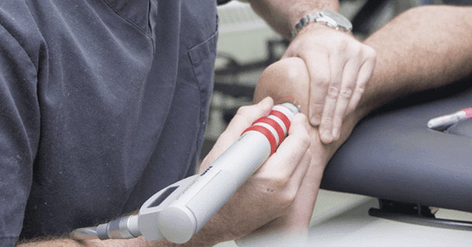
Shock waves are produced by electromagnets creating a signal through water. This signal is is directed through a lens to focus all of the energy created into a single point. This way, the damaged foot is able to receive the full force of the shock wave.
Each shock wave has two potential levels: low energy and high energy. The low energy level of these waves can provide a short-term pain relieving effect, while the high energy level is responsible for stimulating healing. When the high energy pulse comes in contact with the plantar fascia, the body responds by increasing blood flow to the area.
Increased blood flow to damaged or inflamed tissue starts the body’s healing and repair processes. The area receives nutrient-rich blood, and the surrounding tissue begins to produce new, healthy tissue cells to replace the old, damaged ones.
Before this treatment was used specifically for plantar fasciitis, other studies were done on the effects of shock waves for treating wounds and soft tissue pain. These studies proved that shock waves were able to stimulate better wound healing and reduce the number of surgical treatments needed.
Once these studies were established, other researchers decided to focus on studying the healing power of shock waves for painful heel spurs. In these patients, the pain was chronic and unsuccessfully treated with procedures like physical therapy or steroids. During 12-week evaluations, just over 80 percent of the patients said that they noticed complete or almost complete healing, and 17 percent noticed partial improvement.
One study performed one-year evaluations for patients with heel spurs that were treated with high-energy shock waves. 85 percent of patients reported being pain-free and experiencing an improved quality of life.
Two other separate but similar studies also showed that shock waves are effective for treating chronic heel pain associated with plantar fasciitis. In both of these studies, roughly 83 percent of patients reported satisfaction with their treatment.
The studies mentioned above, as well as other studies conducted by the FDA, indicate that this form of therapy is a safe and highly effective treatment for chronic plantar fasciitis both with and without heel spurs.
In every study examined, there are excellent results and no long-term complications. This is important because surgical procedures always carry the risk of nerve damage, infection or additional pain.
If the science is any indication, shock wave therapy be the future of podiatric medicine.