To many people, walking and running seem like some of the most natural processes on earth. However, what many people don’t realize is that the chances are good that they’re going through life with muscular or structural imbalances that can cause injury.
Sedentary people may not necessarily be at risk for injury, but even in the fairly sedentary, these issues – called “biomechanical abnormalities” – can still cause issues.
Gait analysis is often as simple as a trained specialist watching you walk and run. This person is able to look for abnormalities in the way you move. These may include the way your feet tilt, limitation in the range of motion of your ankle, and elevating one side of your pelvis as you step forward.
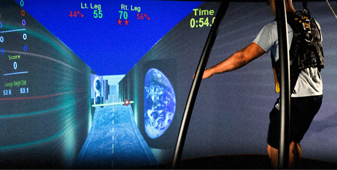
Often, the analyst will film you so they can slow down the video and more precisely look at possible abnormalities. There are also more advanced forms of analysis that involve attaching electrodes to various areas of the body to better map your movement, but these techniques tend to be more costly.
When someone analyzes your gait, they are generally looking for anything that may be causing discomfort or that could contribute to injury. Particularly, they may look for over-pronation or over-supination. Pronation refers to feet that tilt inward, while supination refers to feet that tilt outward. The analyst may also take a brief history of pain to help determine if you have any common issues like Achilles tendinitis or IT band syndrome, or if you are experiencing lower back pain from a gait issue. They also may look fir “hip hitching” which involves over-lifting one side of the hip.
Usually, these issues are caused by muscle imbalances, but they also can be caused by structural issues. Hip hitching, for instance, is often caused by a leg length discrepancy. Sometimes, this can be corrected by using a lift to even out the lengths of the legs. Skilled gait analyst will be able to help you find the best way to address your biomechanical abnormalities.
There are many ways to address biomechanical abnormalities. Depending on the severity of your imbalances, selecting a particular type of shoe may be enough. If you’ve ever been to a sports shoe store, you may have been offered a brief analysis of your gait. The employee can then recommend a typeof shoe that can help correct imbalances. For instance, some shoe manufacturers have a “forefoot cradle” design in some shoes that is essentially an enhanced arch support. This can help to correct overpronation.
If your analysis reveals substantial muscle imbalances, physical therapy may be recommended to help build up muscles that are lacking. Therapy can also help you increase flexibility, like inc cases of ankle equines, which is an issue that inhibits the range of motion in your ankle.
Sometimes, as in the case with some instances of hip hitching, you imbalances may be due to abnormalities in your skeletal structure. In these cases, a doctor or physical therapist might be able to help you fix them via a mechanical intervention like a lift. A lift can be something as simple as a small wedge placed in the bottom of your shoe, and it can help even out your legs and eliminate pelvic tilt issues, at least while you are wearing the shoe.
In short, gait analysis is a simple tool that has the potential to greatly improve your quality of life. Addressing any gait issue preemptively can help you ward off injuries and preserve your general health and well-being.


 Dr. Mikhail Bernshteyn
  Dr. Michael Goynatsky
  Dr. Daniela Escudero
  Dr. Michelle Agyakwah
  Dr. Tatyana Kapustina