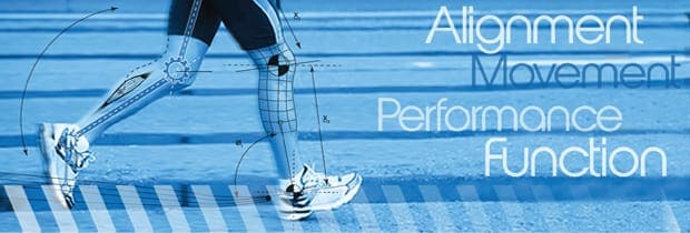
At NYDNR we use most advanced force plate technology and 3D video analysis to evaluate biomechanics of an athlete in motion. This cutting edge analysis is of particular importance to runners and athletes who are involved in cutting and jumping movements.
Biomechanical analysis can register the forces involved with athletic movement. When these forces exceed normal there is excessive stress to joints, muscles and ligaments. The excessive forces are always the result of poor muscle coordination, improper technique (motor control) and muscular weakness. The in depth biomechanical analysis allows differentiation of these factors and provides proper strategy to reduce injury and improve performance.
The combination of force plate and high-speed cameras with intricate software allows us to register and measure:
In order to take the best course of action in a treatment plan for your condition, it’s important to have a keen understanding of the external and internal forces that are at work during movement.
Not only is it important for the staff to understand these forces, it allows you to put your own damage control in place, avoiding further injury or future injuries during movement. Through careful study of biomechanics and motor control, we will be able to help you to
correct your movement patterns. As a result, you can avoid muscle strain, improve shock absorption, avoid overloading your joints and improve performance by becoming more efficient athlete.

Verified Expert Profiles
Dr. Lev Kalika is a world-recognized expert in musculoskeletal medicine. with 20+ years of clinical experience in diagnostic musculoskeletal ultrasonography, rehabilitative sports medicine and conservative orthopedics. In addition to operating his clinical practice in Manhattan, he regularly publishes peer-reviewed research on ultrasound-guided therapies and procedures. He serves as a peer reviewer for Springer Nature.
Dr. Kalika is an esteemed member of multiple professional organizations, including:
Below is a prime example of how ultrasound can take the guesswork out of diagnosis.

A bad physical therapy experience is one of the primary causes of unnecessary surgery
In this instance, an athlete was originally diagnosed with minor quadriceps muscle strain and was treated for four weeks, with unsatisfactory results. When he came to our clinic, the muscle was not healing, and the patients’ muscle tissue had already begun to atrophy.
Upon examination using MSUS, we discovered that he had a full muscle thickness tear that had been overlooked by his previous provider. To mitigate damage and promote healing, surgery should have been performed immediately after the injury occurred. Because of misdiagnosis and inappropriate treatment, the patient now has permanent damage that cannot be corrected.
The most important advantage of Ultrasound over MRI imaging is its ability to zero in on the symptomatic region and obtain imaging, with active participation and feedback from the patient. Using dynamic MSUS, we can see what happens when patients contract their muscles, something that cannot be done with MRI. From a diagnostic perspective, this interaction is invaluable.
Dynamic ultrasonography examination demonstrating
the full thickness tear and already occurring muscle atrophy
due to misdiagnosis and not referring the patient
to proper diagnostic workup
Demonstration of how very small muscle defect is made and revealed
to be a complete tear with muscle contraction
under diagnostic sonography (not possible with MRI)

Complete tear of rectus femoris
with large hematoma (blood)

Separation of muscle ends due to tear elicited
on dynamic sonography examination


