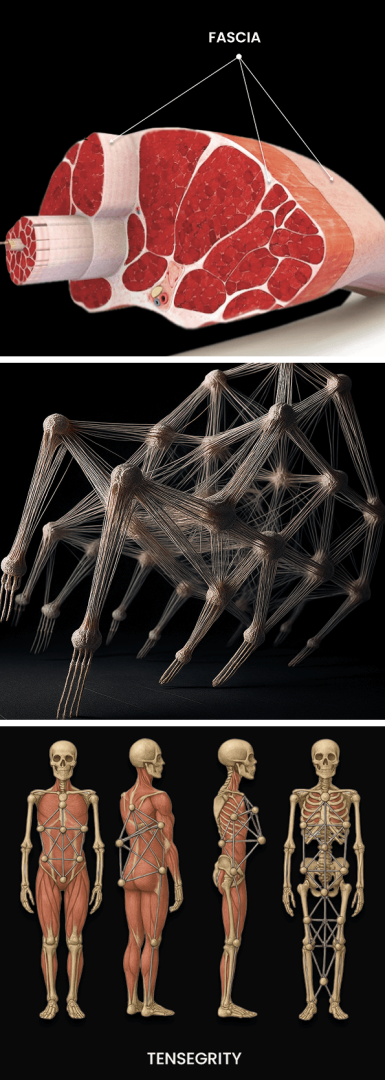
Fascia is a thin, tough network of elastic connective tissue that encases and connects muscles, connective tissues, and internal organs throughout your body, enabling the body’s structures, blood vessels and neural bodies to glide without friction during movement. Until very recently, the role of fascia in biomechanics was poorly understood, and many medical doctors are unaware of its importance in human movement. However, a flourishing body of new research is shedding light on fascia, and how damaged fascia can cause pain and restrict mobility.
The Stecco technique of fascial manipulation is a highly systematic approach that requires specific training. Unlike other manual myofascial therapies, when applied correctly, Stecco Fascial Manipulation not only restores tissue gliding – it reactivates sensory receptors within the fascia, recalibrating the neuromuscular system and restoring efficient motor control.
The Stecco technique also influences the neurohumeral, paracrine, hormonal, and visceral-somatic systems, supporting whole body regulation and recovery. Dr. Kalika has received advanced training directly from Dr. Carla Stecco, creator of the Stecco Method and the world’s leading specialist in fascial science.
At NYDNRehab, we leverage the most advanced, evidence-based technologies, therapeutic approaches, and orthobiologics to restore fascial integrity and enhance biomechanical efficiency, for optimal mobility, stability, and athletic performance.

Clinical director & DC RMSK
Verified Expert Profiles


Orthobiologic, Sports Medicine and Regenerative Medicine Specialist
Fascia plays a vital role in the integration of the body’s various structures and systems. Its impact on the musculoskeletal system and human mobility is one of the greatest discoveries of the 21st Century. Fascia research has become one of the hottest topics in musculoskeletal science, providing evidence-based information that connects the dots of previously poorly understood health phenomena.
Fascia is made up of multiple collagenous layers, lubricated by a gel-like substance called hyaluronan, enabling the tissue to stretch and glide as you move. Fascia is densely embedded with proprioceptors, informing your brain about changes in tissue length, tension, and pressure during dynamic activities. Dense innervation makes fascia capable of generating pain 5X greater than muscle tissue.
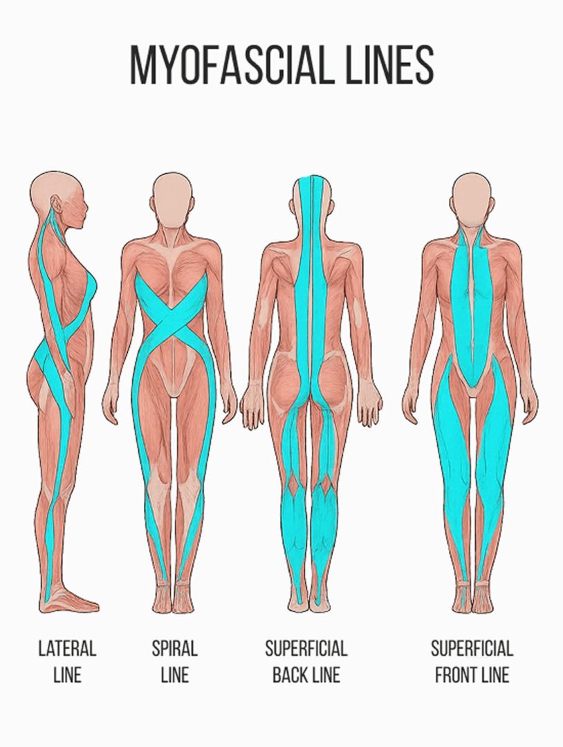
Fascia and muscles work synergistically to support the skeleton, hold organs and structures in place, guide and control movement, and distribute internal and external forces via elastic tension – aka biotensegrity. As long as myofascial tissues maintain their functional properties, biomechanics are optimized for mobility, stability and physical performance.
Biotensegrity can be disrupted when myofascial tissues are injured or damaged in some way. Damaged fascia can become densified and sticky, entrapping nerves and blood vessels and preventing them from gliding among other structures. Densified fascia disrupts muscle action and compromises the elastic tension that controls movement and governs joint alignment.
Factors that disrupt biotensegrity include:
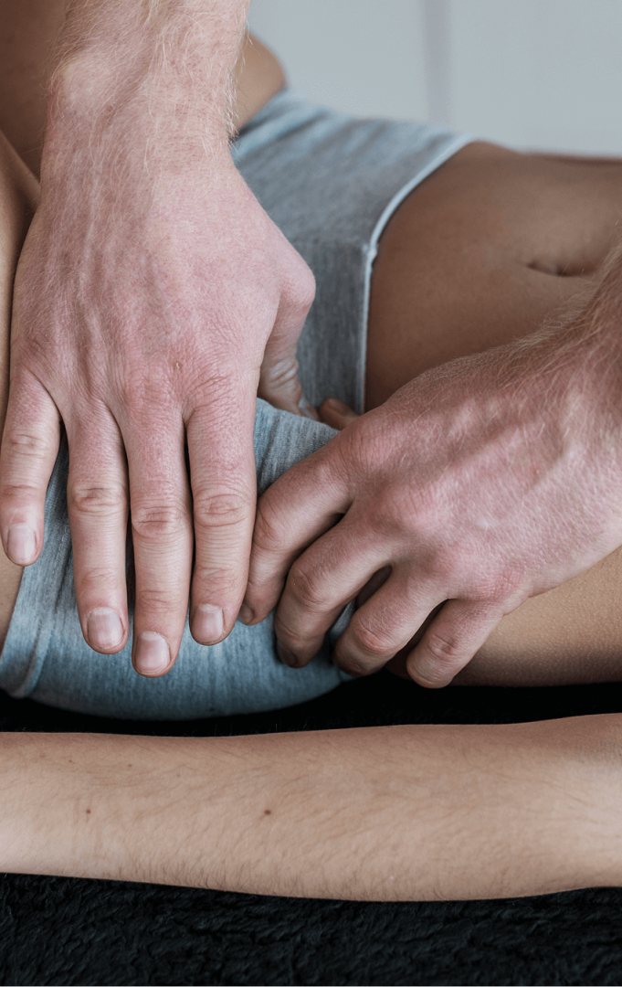
Unhealthy fascia can adhere to other structures, forming adhesions that inhibit mobility and impede muscle action. Fascial manipulation therapy helps to eliminate adhesions and free up densified fascial layers, releasing entrapped nerves and blood vessels, and restoring fascia’s slippery and elastic properties.
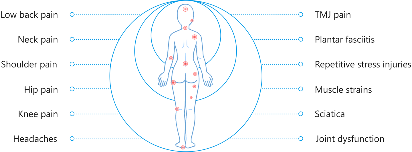
As much as 50% of pain and reduced mobility is due to the erosion of fascial biotensegrity, including loss of elasticity and micro tears in fascial layers. Fascial adhesions are often to blame for pain and restricted movement, but prematurely releasing fascial adhesions prior to restoring biotensegrity can make things worse instead of better.
To restore fascia’s integrity and capacity for force transfer, we first need to restore its elastic properties. Neglecting to properly treat densified or damaged fascia can cause tissue degeneration, leading to serious mobility issues.
Factors contributing to fascial degeneration can be:
Many doctors do not recognize the crucial role of fascia in preventing pain syndromes, movement disorders, and disease, and most have no idea how to correct myofascial dysfunction. Modern doctors are trained to treat pain with medications and steroid injections, without addressing its underlying source. Patients may be referred for physical therapy, but PT alone is not enough to resolve myofascial pain. When pain symptoms persist, surgery may be recommended to repair structural problems that could be more effectively resolved with conservative care.
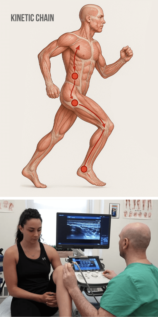
At NYDNRehab, we understand that the body’s systems work together as an integrated whole, and that treating the locus of pain is not enough to eliminate its source. In many cases, pain in one area is referred from mechanical issues farther along the kinetic chain. In the case of myofascial dysfunction, densified fascia can inhibit muscle action, cause joint misalignment, compress and entrap neural bodies, and relay intense pain signals to the brain.
Many massage therapists and physios recognise fascial dysfunction as an important generator of pain and impaired mobility, and many offer fascial manipulation services. Such therapy may provide temporary relief, but without imaging, there is no way to confirm whether biotensegrity has been restored. Superficial densifications and trigger points may be palpable beneath the skin, but deep fascial dysfunction can only be identified and treated with high-resolution ultrasound imaging.
We use dynamic high-resolution ultrasound to explore the myofascial system in real time, to identify areas where biotensegrity has been disrupted. Our patient-centric approach ensures that your treatment plan is customized to fit your unique profile.
Personalized care – we treat the whole patient, not just your symptoms. Our personalized approach ensures your treatment aligns with your unique health profile.
Holistic treatment approach – your body’s structures are interactive and interdependent, designed to work in harmony. Isolating treatment to one area while neglecting the whole body often leads to more pain and dysfunction down the road.
Patient education – transparency and respect make you a stakeholder in your own healing journey. We keep you in the loop during our therapies and procedures, giving you valuable insights and tools for managing your health in the long term.
Cutting-edge tools – our clinic features some of the most advanced technologies and therapies available in rehabilitative medicine. Our therapeutic approach leverages a combination of evidence-based methodologies, for fast and effective results.
Many patients come to us after one or more failed attempts elsewhere, and we are able to deliver results, faster and more effectively. Dr. Kalika applies his expertise in diagnos tic ultrasonography to accurately identify nerve entrapments and resolve fascia-nerve interference syndrome – one of the most common causes of musculoskeletal pain. He has successfully resolved some of the most difficult, chronic, and resistive cases of fascial dysfunction where other practitioners have failed.
The team at NYDNRehab takes a holistic and integrative approach to musculoskeletal disorders. We treat the whole patient, not just your pain symptoms, with the goal of restoring pain-free functional mobility. Dr.Kalika is one of the few practitioners to combine Stecco diagnostic methodology with high resolution diagnostic ultrasonography, to visualize key coordination points of fascial adhesions and tissue thickening.
After reviewing your health history and conducting a comprehensive clinical exam, we use high-resolution ultrasound imaging to dynamically visualize your myofascial system in real time. Your ultrasound exam takes place on-site, on your first visit, where we examine multiple areas of the body in a single session. Our state-of-the-art ultrasound equipment has capabilities for sonoelastography, a tool for testing tissue density and elasticity – key factors in myofascial dysfunction.
Stecco diagnostic methodology combined with ultrasound and sonoelastography lets us:
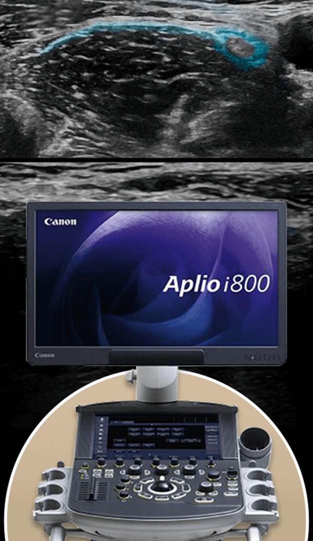
Dr. Kalika is an internationally recognized expert in diagnostic ultrasonography, with multiple research publications to his credit. Unlike conventional imaging modalities such as X-ray or MRI, high-resolution ultrasound provides dynamic images in real time, without radiation and with minimal risk to the patient.
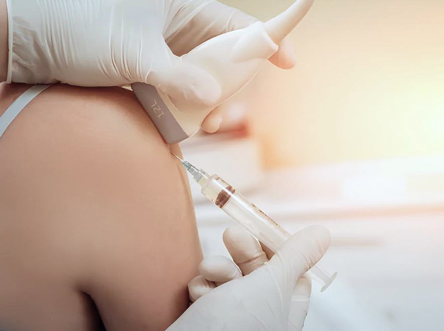
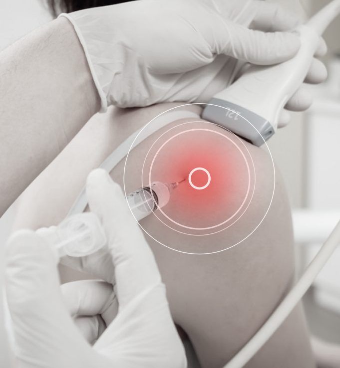
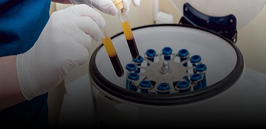
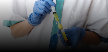
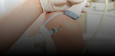
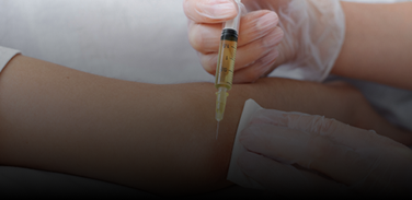
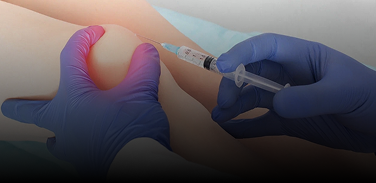
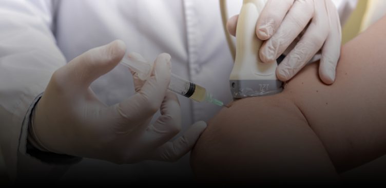
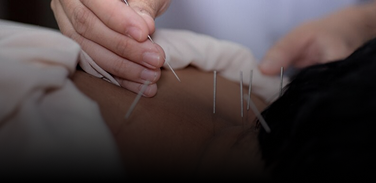
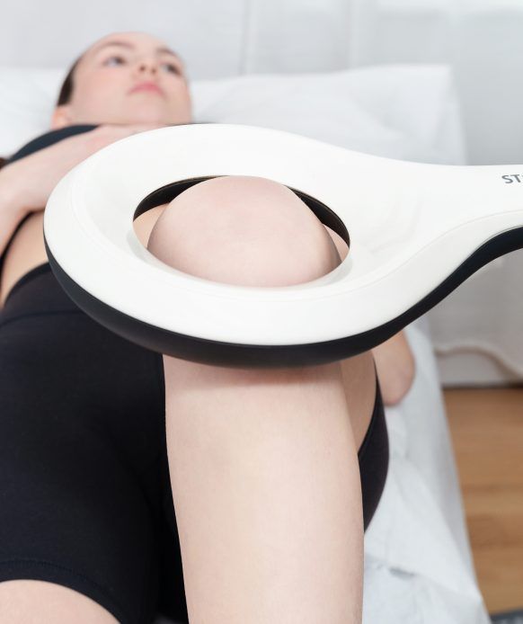
For advanced myofascial release procedures, Dr. Kalika teams up with orthobiologic specialist Dr. Yuri Brogol, combining Stecco fascial manipulation with ultrasound-guided injections of prolotherapy or platelet rich plasma (PRP), and ultrasound-guided hydro release. We use multimodal shockwave therapy and ultrasound-guided dry needling to prepare fascial tissues for treatment, and to stimulate tissue healing.
Dr. Carla Stecco, MD – creator of the Stecco Method of fascial manipulation – is an orthopedic surgeon and professor of human anatomy and movement sciences at the University of Padova in Italy. She has published 180+ peer-reviewed articles and authored several books on the nature of human fascia and its cellular organization. She uses cadaver dissections to educate manual therapists and physicians on the nature and function of fascia in human movement.
Fascia surrounds and connects muscles, engulfs the visceral organs, and anchors organs to muscles, holding them in place during movement. There is a strong relationship between fascia and internal disease that is often overlooked by fascial therapists.
Unlike other manual therapies, the Stecco technique of fascial manipulation is a highly systematic approach that requires specific training. When applied correctly, Stecco Fascial Manipulation not only restores tissue gliding – it reactivates sensory receptors within the fascia, helping the body to reorganize motor control, recalibrating the neuromuscular system and restoring efficient motor control.
Such reorganization occurs not only at the local (receptor and muscle) level, but throughout the entire nervous system, promoting more coordinated and efficient movement patterns. In addition, Stecco Fascial Manipulation influences the neurohumeral, paracrine, hormonal, and visceral-somatic systems, supporting global body regulation and recovery.
Progression of Stecco Fascial Manipulation involves:
These adaptations occur over the course of a week or two as tissue integrity and neural pathways are restored. This advanced methodology works to alleviate pain, eliminate restricted movement, enhance physical performance, and optimize whole-body mobility and stability.
The Stecco Method is the only evidence-based fascial technique that corresponds precisely with fascial science. Stecco methodology should not be confused with other manual therapies such as therapeutic massage, rolfing, Graston Technique, IASMT, or active release techniques. Dr. Kalika is a trained practitioner of the Stecco method.
Fascia manipulation works by targeting fascial restrictions and enhancing tissue mobility. It can be effective for treating certain visceral issues like digestive and reproductive problems. Fascial manipulation can be combined with other therapies to address common musculoskeletal conditions like rotator cuff pain, frozen shoulder, carpal tunnel syndrome, and pelvic disorders.
One recent study compared fascial manipulation to standard physical therapy in 102 patients with low back pain. They found that fascial manipulation significantly impacted scores on the Numeric Pain Rating Scale (NPRS), 15- point Global Rating of Change (GROC), and the Oswestry Disability Index (ODI). Researchers concluded that fascial manipulation has a greater effect on reducing low back pain than standard physical therapy alone.
The superficial fascia of the pelvic floor is a thin fibrous layer of connective tissue embedded with adipocytes – specialized fat cells – along with a complex network of lymphatic and blood vessels. The pelvic fascia plays an important role in exteroception – sensory awareness of stimuli outside the body.
Pelvic pain is mostly generated from hypersensitized nerves embedded in the deep fascia of the pelvis, including the pudendal, gluteal and cluneal nerves that glide among the pelvic floor muscles. Pelvic fascial manipulation is often the missing link in pelvic pain treatment. Pelvic nerves can be released from entrapment by manipulating the endomysium and perimysium. Deep pelvic fascia works synergistically with the transversus abdominis, diaphragm and pelvic floor muscles to optimize abdominal pressure, providing spinal stability. Generous autonomic innervation makes the pelvic fascia sensitive to stress and changes in temperature, and the pelvic fascia is intricately connected to the genitalia.
Other sensory nerves and their endings inhabit the superficial fascia, such as the ilioinguinal, iliohypogastric and genitofemoral nerves. When your parasympathetic nerves are entrapped by superficial fascia they become hyperactivated, generating pelvic pain.
Orthobiologics use natural or neutral solutions, injected under ultrasound guidance to leverage the body’s natural healing mechanisms. The substances work by activating stem cells and growth factors, to stimulate cellular repair and regeneration, and restore fascia’s functional properties.
Orthobiologics are used to treat fascia in a variety of scenarios:

The role of physical therapy is to restore functional mobility in patients presenting with chronic pain syndromes, movement disorders, and acute and chronic injuries. It focuses on strengthening stabilizer muscles and connective tissues, optimizing joint alignment, restoring functional range of motion, and enhancing proprioception. But physical therapy can only be effective if damaged tissues have been pretreated and healed, and are able to manage force loads.
Obstacles to physical therapy success include:
At NYDNRehab, we pretreat damaged tissues to accelerate healing and restore biotensegrity before introducing weight-bearing activities.
Physical therapy can be useful in the later stages of injury treatment, but beginning physical therapy before treating damaged tissues is a recipe for failure that can potentially increase myofascial pain and dysfunction.
Healthy lifestyle factors are key to maintaining fascia that is strong, elastic and resilient. Factors like poor diet and being sedentary can cause fascia to degrade over time, reducing your mobility and increasing your risk of injury.

Fascial pain is often misdiagnosed as muscle pain, and mistreatment can prolong your pain and disability without resolving the problem. At NYDNRehab, we use the highest resolution diagnostic ultrasonography to visualize your fascia in motion, in real time.
Once we identify the location and scope of fascial tissue dysfunction, we develop a personalized plan for restoring fascia integrity, using the most advanced technologies and evidence-based methodologies available.
Don’t settle for ineffective treatments that only target pain symptoms without providing lasting solutions. Contact NYDNRehab today for advanced holistic therapy that gets to the root cause of your myofascial pain and eliminates it for good.


Research authored or co-authored by the clinic’s medical director. The following research publications inform the clinical approach used in this treatment program.
Scientific abstract
2021
 Lev Kalika
  Rostyslav Bubnov
 Conference abstract
2022
Lev Kalika Rostyslav Bubnov 
Below is a prime example of how ultrasound can take the guesswork out of diagnosis.

A bad physical therapy experience is one of the primary causes of unnecessary surgery
In this instance, an athlete was originally diagnosed with minor quadriceps muscle strain and was treated for four weeks, with unsatisfactory results. When he came to our clinic, the muscle was not healing, and the patients’ muscle tissue had already begun to atrophy.
Upon examination using MSUS, we discovered that he had a full muscle thickness tear that had been overlooked by his previous provider. To mitigate damage and promote healing, surgery should have been performed immediately after the injury occurred. Because of misdiagnosis and inappropriate treatment, the patient now has permanent damage that cannot be corrected.
The most important advantage of Ultrasound over MRI imaging is its ability to zero in on the symptomatic region and obtain imaging, with active participation and feedback from the patient. Using dynamic MSUS, we can see what happens when patients contract their muscles, something that cannot be done with MRI. From a diagnostic perspective, this interaction is invaluable.
Dynamic ultrasonography examination demonstrating
the full thickness tear and already occurring muscle atrophy
due to misdiagnosis and not referring the patient
to proper diagnostic workup
Demonstration of how very small muscle defect is made and revealed
to be a complete tear with muscle contraction
under diagnostic sonography (not possible with MRI)

Complete tear of rectus femoris
with large hematoma (blood)

Separation of muscle ends due to tear elicited
on dynamic sonography examination


