Ultrasound Guided Procedures

In orthopedics and sports medicine, orthobiologic injection procedures and regenerative therapies are frequently used to reduce pain and inflammation, and to stimulate healing of injured or damaged tissues by attracting agents like stem cells and growth factors to the treatment site. Musculoskeletal injuries typically affect multiple tissue types, and each type has unique treatment requirements. Without clear imaging it is impossible to precisely target diverse tissue types with injections, shockwaves and other therapies, especially when treating deep tissues.

At NYDNRehab, high resolution ultrasonography provides a safe and effective imaging solution for various rehabilitative procedures, allowing for precise and effective treatment that speeds up tissue healing.

Contact us »

We’re located on 25th street in Manhattan NYC.
Open Monday-Friday, 10am-8pm.

Dr. Lev Kalika

Clinical director & DC RMSK

About Holistic Healing Experts Dr. Kalika and Dr. Brosgol

Dr. Lev Kalika, DC clinical director of NYDNRehab, is an internationally recognized expert in diagnostic and musculoskeletal ultrasound imaging, with multiple research papers to his credit. Dr. Kalika has studied with some of the world’s most prestigious experts in diagnostic, fascia, and nerve ultrasonography, and has presented his research at multiple international professional conferences. Dr. Kalika is an active member of the American Institute of Ultrasound in Medicine (AIUM), and has developed his own unique approach to Dynamic Functional and Fascial Ultrasonography.
Dr. Yuri Brosgol

Orthobiologic specialist

Dr. Yuri Brosgol, MD is a neurologist with 20+ years of experience in treating pediatric and adult myofascial pain. When emerging research on the critical role of fascia in human mobility captured Dr. Brosgol’s interest, he pursued training in orthobiologics and fascial release techniques. Dr. Brosgol learned fascial hydro release techniques directly from Dr. Carla Stecco, the world’s leading specialist in fascial science.

Dr. Brosgol has become a pioneer in the use of orthobiologic solutions, paving the way for transformative advancements in the treatment of myofascial disorders.

Together, Dr. Kalika and Dr. Brosgol are combining their skills to revolutionize the way musculoskeletal pain and injuries are treated. Dr. Kalika’s successful track record of rehabilitating musculoskeletal injuries combined with Dr. Brosgol’s expertise in treating myofascial pain makes NYDNRehab the clinic of choice for injury rehab in NYC.

icon

How Musculoskeletal Injuries Become Chronic,
and Why Mainstream Doctors Can't Fix Them

Everyone suffers minor injuries from time to time, and many of them self-resolve over time with rest and basic first aid. But some injuries get worse instead of better, and many patients wait until their symptoms become unbearable before they seek professional intervention.

Finding the right doctor is often a conundrum for people who don’t look beyond conventional medicine. It is not uncommon for patients with pain syndromes and movement disorders to go from one practitioner to another, to be subjected to a plethora of treatment approaches, with no enduring results. In the process, the patient suffers inconvenience, wastes valuable time, and depletes their bank account, all while their health and mobility continue to deteriorate.

The goal of rehabilitative medicine is to restore pain-free functional movement and enhance the patient’s quality of life. But the truth is that the vast majority of practitioners have no idea how to approach or resolve chronic pain and dysfunction. Conventional medicine focuses on pain management, prescribing drugs and steroid injections that temporarily mask pain symptoms, without improving mobility. Patients may be referred for physical therapy, but physical therapy alone is not enough to resolve motor dysfunction.

When it comes to physical therapy, there are multiple factors that can limit its effectiveness:

  • Pain, even while managed with drugs
  • Tissue damage that affects mechanoreceptors embedded in joints and fascia, impairing kinesthetic awareness necessary for good mechanics
  • Misdiagnosis or underdiagnosis leading to inappropriate or ineffective treatment that prolongs patient suffering
  • Dogmatic healthcare approaches that rely on ineffective exercises and obsolete or unproven therapies
  • Unnecessary invasive interventions and surgeries
  • Tunnel vision that treats symptoms but neglects the whole patient
  • Ignorance of, or lack of exposure to emerging evidence-based approaches that really work
  • Lack of teamwork among various practitioners

In order for physical therapy to provide effective and lasting results, we must first remove obstacles and restore biotensegrity – the optimal state of mechanical integrity governed by the myofascial system. That means addressing structural issues that affect multiple systems, including organs and functional units made up of fascia, muscles, tendons, ligaments, joints, nerves and blood vessels. In addition, the patient’s emotional health can present an obstacle to successful rehabilitation.

At NYDNRehab, your rehabilitation journey begins with pre-treating damaged and dysfunctional tissues to eliminate pain and inflammation and restore biotensegrity to the body’s systems. Advanced orthobiologic injection therapies play a critical role in injury rehab that cannot be filled by physical therapy alone.

The Important Role of Fascia in
Biotensegrity

Conventional medicine takes a reductionist approach to injury treatment, zeroing in on the locus of pain while ignoring more distal structures. By contrast, holistic medicine considers the interdependent nature of the entire organism, exploring how damage or dysfunction in one area of the body is related to pain symptoms in another.

Fascia is a complex web-like network of connective tissue that surrounds and connects muscles, engulfs the visceral organs, and anchors organs to other structures, holding them in place during movement. Fascial tissue is thin, tough and elastic, able to stretch and glide as you move. It is made up of collagen fibers, lubricated by hyaluronic acid – a slippery gel-like substance able to attract 100X its mass in water.

Until recently, the role and significance of fascia was poorly understood, if not completely disregarded. In fact, in cadaver labs used for research, fascia is often cast aside as unnecessary waste. But over the past decade, volumes of research have emerged that recognize the critical role of fascia in human movement.

Fascia is fundamental to human mobility and stability:

  • Fascia encases the muscle, providing a nourishing environment for muscle performance
  • Fascia covers the abdominal and urogenital viscera and anchors them in place
  • Fascia covers and protects the bones and blood vessels

When fascia is overworked or depleted due to trauma, mechanical stress, or lack of fluids and nutrients, it can become dehydrated and fibrous. Trigger points can form in fascia and muscle tissue, causing pain and interfering with muscle function. Damaged fascia can become dense and sticky, creating friction and impeding the ability of nerves and blood vessels to glide among other structures. Densified fascia disrupts muscle coordination patterns, causing reduced performance and increased risk of injury.

Fascia is generously embedded with mechanoreceptors that inform your brain of your body’s position as you move. When fascia is injured, proprioception becomes impaired, reducing movement efficiency and increasing your risk of injury. There is a strong relationship between fascial integrity and internal disease that is often overlooked by medical doctors and fascial therapists.

During physical activity, fascia works together with muscle to provide tensile integrity – aka biotensegrity – to guide and control movement, hold organs and other structures in place, and mediate and distribute outside forces. When muscles, fascia or both are damaged, biotensegrity is compromised and mobility is impaired.

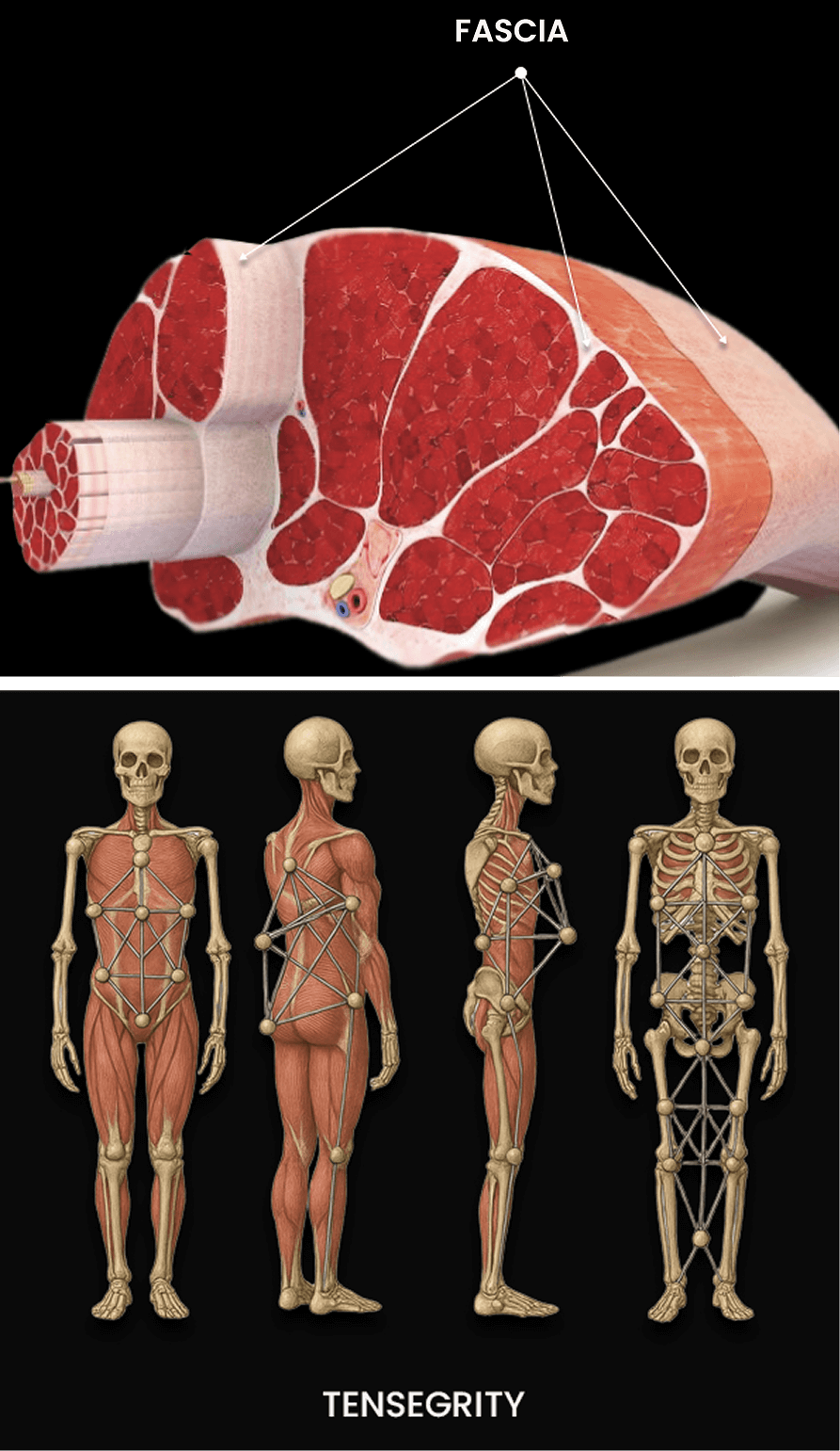
Restoring biotensegrity is a key factor in injury rehabilitation. It is not enough for injured muscles tissues to heal and pain to subside. Unless the damaged fascial layers are treated, mobility will continue to be impaired, increasing the risk of future injuries.

At NYDNRehab, we use the following interventions to restore fascia’s slippery and elastic properties:

1
Ultrasound guided interfascial plane and nerve hydrodissection, an injection procedure that "unsticks" fascial layers and liberates nerves and blood vessels.
2
Stecco myofascial release, an evidence-based manual approach to restoring fascial integrity.
3
Ultrasound guided extracorporeal shockwave therapy, to realign collagen fibers, promote fascial hydration, and restore tissue gliding.
4
Orthobiologic injection therapies to promote collagen production and eliminate fascial densifications and adhesions.

Our fascia-first approach to injury rehab lays the groundwork for optimal mobility and enhanced physical performance.

Personalized Advanced Therapy Brings Fast and Enduring Results

Many run-of-the-mill physical therapy clinics rely on symptoms-based diagnosis, antiquated recovery timelines, and cookie-cutter exercise regimens that are not up to speed with the most current evidence. Patients are often released with only small improvements in mobility, resigned to adapting to a “new normal.”

For athletes, the stakes are even higher. When released back to sport with injuries that are not fully rehabilitated, reinjury risk is dramatically elevated and performance is often impaired compared to pre-injury metrics. The decision to release an athlete back to play is often based on recovery timelines for a particular injury. Once that time has elapsed, pressure from coaches and parents, and even the athlete, can lead to premature release that sets the patient up for failure.

At NYDNRehab, our goal is to fully restore functional pain-free mobility that meets or exceeds the patient’s pre-injury condition. Patient diagnosis, treatment and release are based on our extensive experience and expertise. Our decision-making is backed by scientific evidence, objective data, and expertise in a broad range of regenerative and advanced methodologies.

Our personalized one-on-one therapy sessions mean that your treatment protocol is tailored-made to address the unique characteristics of your injury and your unique anatomy. Your release date is based on a checklist of performance parameters derived from quantitative analysis, and confirmed by ultrasound imaging.

icon

Our Ultrasound Guided Orthobiologic Procedures

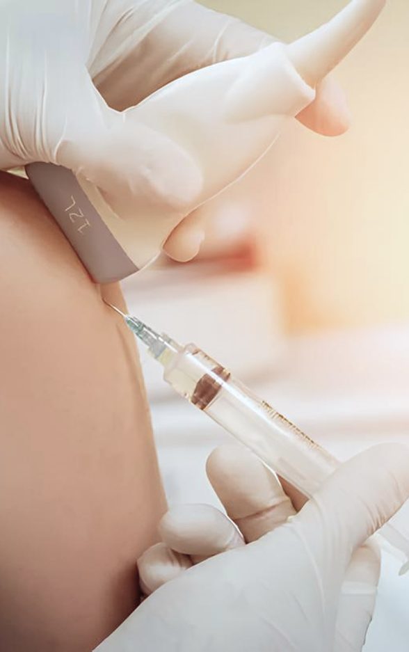
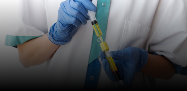
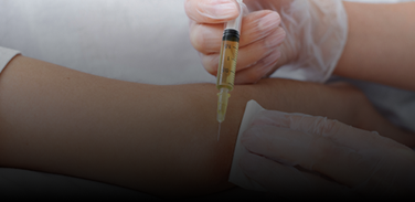
Orthobiologic injection therapies use natural/neutral solutions, injected with precision thanks to ultrasound guidance. The injected solutions stimulate cellular repair by either nourishing or irritating the targeted cells. Needling procedures like dry needling and PENS use filament-thin non-medicated needles to target myofascial trigger points and normalize neural activity..

For ultrasound-guided needling procedures, Dr. Kalika partners with orthobiologic specialist Dr. Brosgol to ensure the needles hit their mark. Treatment results are dramatically enhanced when combined with focused extracorporeal shockwave therapy (fESWT), another area of expertise for Dr. Kalika.

Orthobiologic procedures available at NYDNRehab include:

Platelet Rich Plasma (PRP)

PRP therapy uses a sample of the patient’s own whole blood, spun in a centrifuge to extract a high concentration of platelets. When injected into damaged tissues, PRP initiates tissue repair by releasing biologically active agents such as growth factors, cytokines, lysosomes and adhesion proteins. To be effective, it is critical to use the right concentration and quality of platelets, and to and follow proper isolation techniques. When administered correctly, PRP can help to jump-start tissue healing in chronic injuries and accelerate repair in acute injuries.

Matrix PRP

For tendon ruptures, Matrix PRP takes PRP therapy to the next level by creating a collagenous bridge between the walls of the tear and the rest of the tendon. Matrix is a highly concentrated PRP, diluted and mixed with fibrinogen. At the injection site, the solution becomes a gel-like collagenous substance that adheres to the walls of the tear and fills the space between them, providing a scaffold for platelets to better deliver growth factors and accelerate tissue healing. The fibrin matrix helps to stabilize growth factors and attract stem cell migration to the treatment site.

Platelet Releasate Therapy

Platelet releasate therapy involves injecting platelet releasate – a mixture of growth factors and biomolecules – into injured muscles and tendons to promote healing. Platelet releasate works by activating leukocytes and endothelial cells, and stimulating blood vessel growth.

Alpha-2-Macroglobulin (A2M)

Alpha 2 macroglobulin (A2M) is a naturally occurring blood plasma protein that acts as a carrier for numerous proteins and growth factors. As a protease inhibitor, A2M reduces inflammation in arthritic joints and helps to deactivate a variety of proteinases that typically degrade cartilage.

Prolotherapy and Prolozone

Prolotherapy uses a biologically neutral solution to irritate stubborn tissues, triggering the body’s innate healing mechanisms to grow new normal tendon, ligament and muscle fibers. Prolotherapy is often used for slow-to-heal tendon and ligament ruptures, where low vascularity inhibits tissue healing.

Prolozone takes Prolotherapy to the next level by adding a combination of procaine, anti-inflammatory medications, vitamins, and minerals, followed by a mixture of ozone/oxygen gas, injected into targeted joints or tissues. When performed under ultrasound guidance, Prolozone therapy quickly reduces pain and inflammation while jump-starting the healing process.

Hyaluronic Acid Injections

Hyaluronic acid is a natural component of joint synovial fluid. Its slippery gel-like properties provide lubrication that reduces friction, enabling joints, muscles and fascia to move freely without pain. In arthritic patients, synovial hyaluronic acid tends to break down, causing bony structures to rub against one another. Hyaluronic acid is also a primary component of fascial tissue, providing its slippery gliding properties. Hyaluronic acid injections can help to relieve joint and myofascial pain by eliminating friction and restoring pain-free mobility.

Hyaluronic acid is a natural component of joint synovial fluid. Its slippery gel-like properties provide lubrication that reduces friction, enabling joints, muscles and fascia to move freely without pain. In arthritic patients, synovial hyaluronic acid tends to break down, causing bony structures to rub against one another. Hyaluronic acid is also a primary component of fascial tissue, providing its slippery gliding properties. Hyaluronic acid injections can help to relieve joint and myofascial pain by eliminating friction and restoring pain-free mobility.

Injuries often involve fascial tissue that has thickened and become sticky, often adhering to other structures. In the process, nerves and blood vessels can become entrapped, causing pain and restricting mobility. The hydrodissection procedure injects a saline solution into densified fascial layers under ultrasound guidance, separating the layers and releasing entrapped nerves and blood vessels. Hydrodissection is often used in conjunction with manual fascial manipulation, to restore fascial integrity.

Ultrasound Guidance Makes Our Injection Therapies More Accurate and Effective

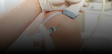
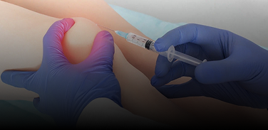
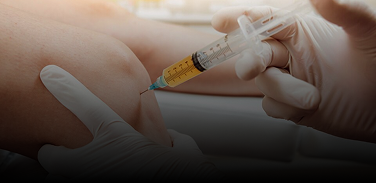
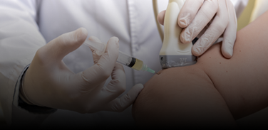
Without ultrasound imaging, therapeutic procedures are hit-or-miss, often failing to achieve their goals. Guidance by high resolution ultrasound ensures that injected solutions reach their intended tissues, without bleeding over into other structures. This means faster pain relief and accelerated healing, often with fewer treatment sessions.

During needling procedures, ultrasound guidance protects nerves and blood vessels from accidental needle penetration while ensuring that injected substances hit their target. Research shows that ultrasound guidance of needling procedures results in superior patient outcomes in terms of pain relief and enhanced mobility.

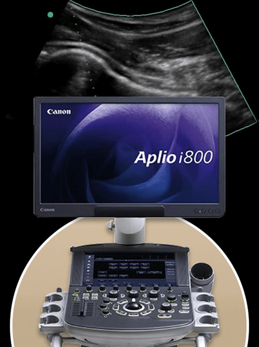
It is important to note that only advanced high resolution ultrasound shows us minute details that cannot be seen with regular ultrasound imaging. High resolution imaging is critical for fascial and nerve injections, and for treating tendon tears.

Explore Dr. Kalika's Published Research on
Ultrasound Guided Procedures

Dr. Kalika and his research colleague, Dr. Rostyslav Bubnov, have conducted extensive research on the use of high resolution ultrasound imaging in the treatment of a broad range of conditions.

Following is a small sampling of Dr. Kalika’s work:

1
A 2022 study published in Neuromodulation explores the efficacy of ultrasound-guided focused, defocused and radial shockwave therapy in treating low back pain.
2
A 2023 study published in Continence demonstrated the effect of ultrasound-guided dry needling in treating patients with myofascial pelvic pain.
3
A 2024 randomized controlled trial supported the use of ultrasound-guided dry needling to manage neck pain and vertigo.

You can gain access to more of Dr. Kalika’s research by following this link

icon

Get the Best High Resolutions Ultrasound Guided Therapy in NYC

There are dozens of physical therapy clinics in Manhattan, but none of them can compete with the expertise and cutting edge technologies featured at NYDNRehab. Our dedication to holistic solutions that bring fast and effective results, along with our patient-first approach and personalized treatment plans, make NYDNRehab the premiere Manhattan center for physical rehabilitation.

Don’t waste your time and money on one-size-fits-all solutions that fail to resolve your condition. Contact NYDNRehab today, and see how personalized holistic therapy can help you rediscover what it’s like to move confidently, without pain or limitations.

Our Awards

Clinical Case Studies
NYDNRehab


Case Study: Chronic Right-Sided Neck Pain after Dry Needling

Our Patient Our patient is a female in her 20s who came to us with severe right-sided neck pain, consequent to a dry needling session at another clinic. Her pain spanned from the upper trapezius area and down the medial scapular border. The patient has a history of chronic right-sided neck pain and previously had […]

learn more

Case Study: Lateral Epicondylitis

Our Patient Our patient is a 56 year-old male jeweler who came to us with lateral elbow pain. The patient is a very athletic tennis player who works out at the gym on a regular basis. He stated that his pain initially started at the front of his elbow and eventually moved toward his lateral […]

learn more

Ultrasound Guided Procedures FAQs

Is ultrasound imaging safe for everyone?
Ultrasound is the safest imaging modality available – safe enough to visualize your unborn baby in the womb. By contrast, imaging tools like Xray and MRI expose patients to harmful radiation, making them unsuitable for vulnerable populations and people with implanted medical devices, such as pacemakers.
What makes ultrasound imaging different from MRI?
Ultrasound has multiple advantages over MRI:
  • Dynamic imaging allows us to visualize the patient in motion, in real time
  • Convenient on-site imaging saves time, with no waiting for lab results
  • Ultrasound is dramatically more comfortable for the patient, with no need to change clothes or hold perfectly still
  • We can visualize multiple sites in the same session, to compare different sides of the body, trace the path of long structures like muscles, bones and nerves, and elicit patient feedback throughout the session for greater accuracy
  • Ultrasound is much less expensive for both doctor and patient
  • Ultrasound equipment is mobile, with minimal space requirements
How does ultrasound guidance support injection procedures?
Needling procedures often target tiny spaces that are impossible to palpate or pinpoint, such as joint capsules, deep tissue trigger points, and scar tissues. Needles can accidentally penetrate blood vessels and nerves, and injections can completely miss their mark, spilling into healthy tissues. Ultrasound guidance helps to avoid accidents and ensures that the procedure precisely hits its intended target.
Do I need to go to a special lab for ultrasound imaging?
Most medical doctors and physical therapists do not have the skills or equipment to do ultrasonography on-site. Dr. Kalika is one of a handful of practitioners in the USA with the training and expertise to accurately perform diagnostic ultrasound. We use highest-resolution ultrasound for diagnosis and guidance. Your diagnostic ultrasound exam takes place on your first visit, in the comfort of our Manhattan clinic.

    Get Advanced Holistic Therapy

    Provide your contact details and we will contact you during our operating hours


    Research at NYDNRehab

    Bubnov, Rostyslav, Lev Kalika, and Zbigniew Pilecki. "P181 ULTRASOUND-GUIDED DRY NEEDLING FOR VERTIGO MANAGEMENT IN A COMBAT PATIENT WITH SKULL TRAUMA: A COMPREHENSIVE CASE STUDY." Neuromodulation: Technology at the Neural Interface 28.1 (2025): S276.
    Kalika, Lev, and Rostyslav Bubnov. "PO236/# 817 TARGETED ULTRASOUND-GUIDED SHOCKWAVE THERAPY OF LOW BACK PAIN USING FOCUSED, DEFOCUSED AND RADIAL SHOCKWAVE: E-POSTER VIEWING." Neuromodulation 25.7 (2022): S339.
    Ultrasound Guided Dry Needling for Acute and Chronic Low Back Pain Imaging and Clinical PatternsUltrasound Guided Dry Needling for Acute and Chronic Low Back Pain Imaging
    Bubnov, R., et al. "EFFECT OF ULTRASOUND-GUIDED DRY NEEDLING ON PELVIC PAIN, BLADDER NECK MOTILITY, AND POSTURAL IMBALANCE IN PATIENTS WITH MYOFASCIAL PELVIC PAIN SYNDROME: A MULTILEVEL MULTIPARAMETER NEUROMUSCULAR ULTRASOUND STUDY." Continence 7 (2023): 100813.
    Lee, Jong H., Jae U. Lee, and Seung W. Yoo. "Accuracy and efficacy of ultrasound‐guided pes anserinus bursa injection." Journal of Clinical Ultrasound 47.2 (2019): 77-82.
    bg

    In this instance, an athlete was originally diagnosed with minor quadriceps muscle strain and was treated for four weeks, with unsatisfactory results. When he came to our clinic, the muscle was not healing, and the patients’ muscle tissue had already begun to atrophy.

    Upon examination using MSUS, we discovered that he had a full muscle thickness tear that had been overlooked by his previous provider. To mitigate damage and promote healing, surgery should have been performed immediately after the injury occurred. Because of misdiagnosis and inappropriate treatment, the patient now has permanent damage that cannot be corrected.

    The most important advantage of Ultrasound over MRI imaging is its ability to zero in on the symptomatic region and obtain imaging, with active participation and feedback from the patient. Using dynamic MSUS, we can see what happens when patients contract their muscles, something that cannot be done with MRI. From a diagnostic perspective, this interaction is invaluable.

    Dynamic ultrasonography examination demonstrating
    the full thickness tear and already occurring muscle atrophy
    due to misdiagnosis and not referring the patient
    to proper diagnostic workup

    Demonstration of how very small muscle defect is made and revealed
    to be a complete tear with muscle contraction
    under diagnostic sonography (not possible with MRI)

    image

    Complete tear of rectus femoris
    with large hematoma (blood)

    image

    Separation of muscle ends due to tear elicited
    on dynamic sonography examination

    bg
    Buy now 3D Gait
    Payment Success
    Request Telehealth Request Telehealth Request in office visit Book now