Clinical director & DC RMSK
Verified Expert Profiles
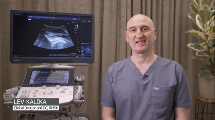
Dr. Lev Kalika, DC clinical director of NYDNRehab, is an internationally recognized expert in diagnostic and musculoskeletal ultrasound imaging, with multiple research papers to his credit. Dr. Kalika has studied with some of the world’s most prestigious experts in diagnostic, fascia, and nerve ultrasonography, and has presented his research at multiple international professional conferences.
Lev Kalika has refined his approach to knee osteoarthritis treatment by introducing cutting-edge therapies and methodologies that go beyond pain management to halting and reversing cartilage degeneration. His expertise in diagnostic ultrasonography ensures that every patient receives personalized treatment, based on high-resolution imaging.
Dr. Kalika is an active member of the American Institute of Ultrasound in Medicine (AIUM), and has developed his own unique approach to Dynamic Functional and Fascial Ultrasonography.

Orthobiologic, Sports Medicine and Regenerative Medicine Specialist
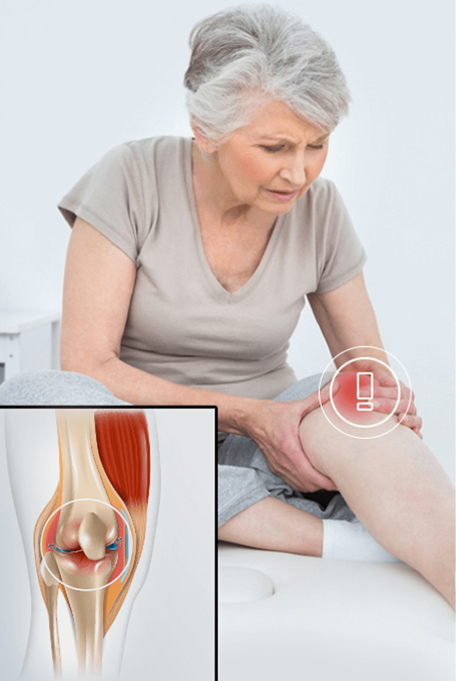
Conventional treatment for knee OA is geared to pain management with drugs and corticosteroid injections. Physical activity is discouraged, leading to further tissue degeneration, until surgery becomes the only option. While knee replacement surgery can provide an effective solution, it is risky, costly, and often unnecessary.
Multiple studies have proven that in most people, the primary causes of joint degeneration are sedentary lifestyle and obesity. In fact, one analysis revealed that the prevalence of knee osteoarthritis has more than doubled since the mid-20th century due to lifestyle factors, and another peer-reviewed article debunks the notion that “wear and tear” is the underlying cause of joint OA.
At NYDNRehab, our holistic approach to knee OA goes beyond pain management, to finding solutions that halt cartilage degradation and restore the knee’s structural integrity. Factors like muscle imbalances, poor joint alignment, damaged fascia, trigger points and nerve entrapment can all affect knee mechanics, creating dysfunction that worsens your condition. Our personalized approach to patient care ensures that all contributing factors are detected and addressed, so you can regain functional mobility.
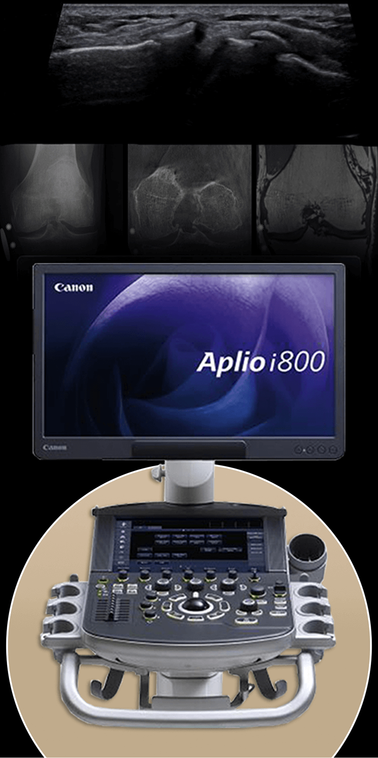
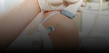
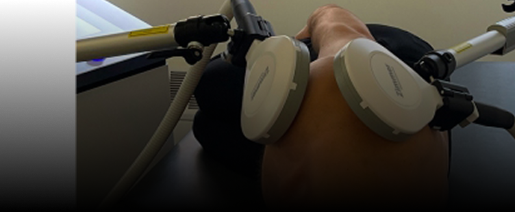
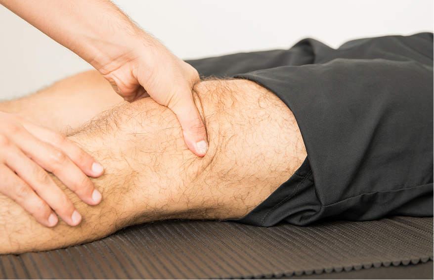
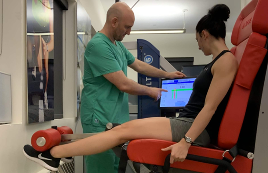
We use high resolution ultrasonography to dynamically visualize your knee in real time, to determine whether structural (anatomical) changes are causing pain and dysfunction, or if dysfunction is causing structural changes. Your comprehensive ultrasound exam takes place on-site, on your first visit, so we can start your healing journey right away.
Dr. Kalika’s expertise in diagnostic ultrasonography enables him to visualize your knee in motion, to identify the exact location where cartilage has eroded, and to see how OA impacts your joint function. Compared to MRI, where knee mechanical issues rarely present themselves, diagnostic ultrasonography has a resolution up to 100X higher than MRI for visualizing superficial tissues. Dynamic high-resolution imaging gives us a full picture of the knee and its supporting structures as they interact, in real time.
Ultrasound surpasses MRI as a diagnostic tool in multiple ways:
In addition to ultrasound imaging, our high-tech 3D gait analysis lab lets us identify inefficient gait mechanics that contribute to knee OA pain. We not only treat your knee OA, but we correct your gait, realign your joints, improve your posture, and strengthen supporting structures to promote stability and minimize knee joint wear-and-tear.
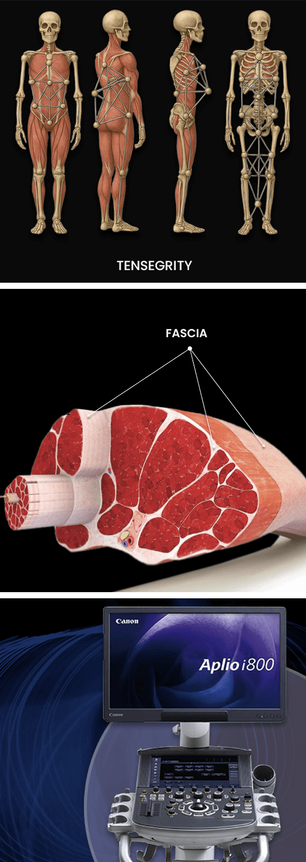
Knee degeneration often begins with overload due to knee instability and inefficient gait, gradually affecting knee mechanics. Multiple structures converge at the knee, supporting its capacity to manage force loads many times your body weight. But muscle weakness and imbalances, poor joint alignment, and other factors can undermine knee function. To stop the degenerative process, we need to address the factors that affect knee stability, beginning with biotensegrity.
Biotensegrity refers to tensile integrity – a state where a system of individual components is held together under continuous elastic tension. In the human body, biotensegrity is created by the myofascial system, the network of muscles, fascia, and nerves that work together to produce, control, and guide forces, and to hold the body’s various organs and structures in place as you move.
Biotensegrity can be disrupted when myofascial tissues are injured or damaged in some way. When that happens, nerves and blood vessels can become entrapped in densified fascial tissue, preventing them from gliding among other structures and producing pain. At the same time, the elastic tension that governs joint alignment and controls movement is disrupted, creating motor deficits that undermine mobility and stability.
Factors that disrupt myofascial biotensegrity include:
Many doctors do not understand the crucial role of the myofascial system in preventing pain syndromes, movement disorders, and disease. In fact, most medical doctors have no idea how to correct myofascial dysfunction or even recognize it as a factor. They simply treat pain symptoms with medications and eventually recommend surgery.
At NYDNRehab, we understand that the body’s systems work together as an integrated whole, and that treating pain is not enough to eliminate its source. We use dynamic high-resolution ultrasound to explore the myofascial system in real time. Ultrasound imaging lets us visualize muscles, fascia, nerves and other structures in motion, to identify places where biotensegrity has been disrupted.
Once we identify the problem, we use the most advanced therapeutic approaches to restore myofascial integrity and promote tissue healing.
At NYDNRehab, we pull out all the stops to halt knee pain and inflammation, and restore pain-free mobility. Our holistic multimodal approach is based on the most current scientific evidence. Our advanced methodologies go beyond pain management, to actually regenerating new cartilage and restoring knee function.
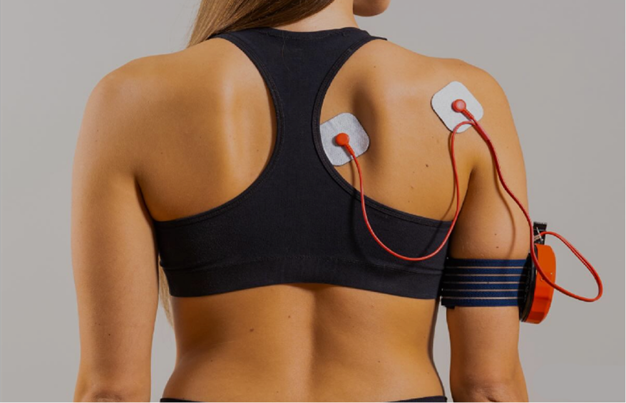
Prior to beginning physical therapy, we pretreat your knee tissues to reduce inflammation and stimulate cell regeneration. Pretreatment addresses pain and inflammation, and releases entrapped nerves – a factor that accounts for 50% of pain and dysfunction in knee OA cases
Obstacles to physical therapy success include:
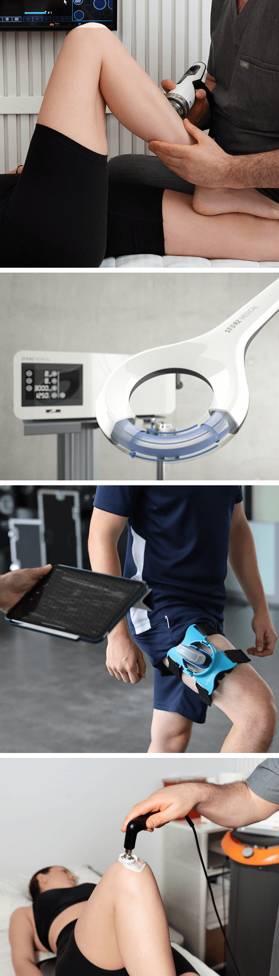
At NYDNRehab, we use a broad range of regenerative technologies, orthobiologics, and integrative therapeutic approaches to resolve issues that can stand in the way of successful physical therapy. Our staff is certified in a diverse array of holistic treatment methodologies, and our one-on-one treatment sessions are personalized, based on your unique diagnostic profile.
Without pretreatment, physical therapy is doomed to fail, and exercise may even do more damage to already compromised structures. Once your knees are ready to tolerate loads, we customize a physical therapy plan to restore strength, stability and range of motion. Your one-on-one personalized physical therapy sessions are designed to help you achieve the best results in the least amount of time.
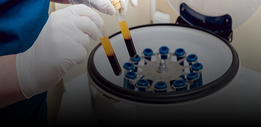
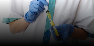
Conventional orthopedists may inject PRP or other solutions to ease the symptoms of knee OA, but orthobiologic injections alone are not sufficient to restore pain-free knee mobility. At NYDNRehab, our integrative approach goes beyond pain management – our goal is to optimize knee mobility, stability and function.
Our holistic integrative approach to knee OA begins with prehab:
We follow a complex process to optimize orthobiologic results:
Our comprehensive integrated approach combines the healing power of orthobiologics with advanced methodologies for strengthening, balancing and stabilizing the body’s structures. The combined expertise of doctors Kalika and Brosgol ensure that you get the very best care available for treating and resolving knee OA.
Unless you are 100% certain you need a knee replacement, you don’t need to see an orthopedic surgeon to treat your knee OA. Early and mid-stage knee OA is best treated with conservative holistic approaches that incorporate advanced methodologies.
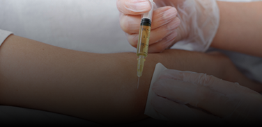
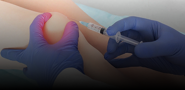
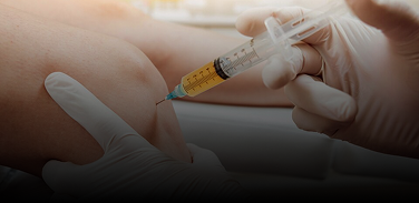
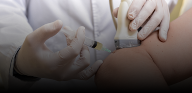
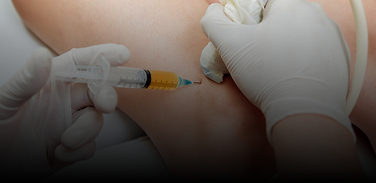
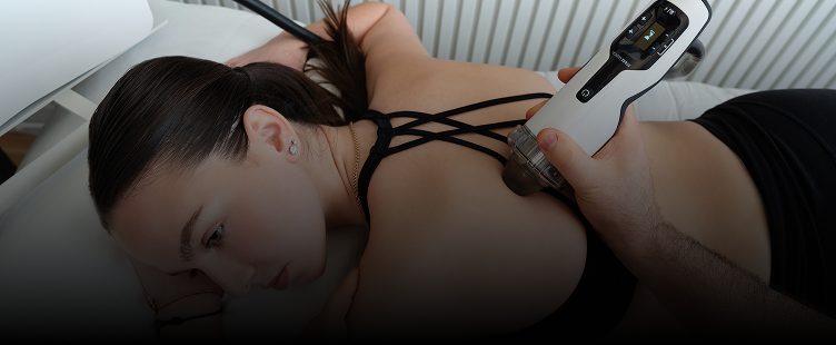
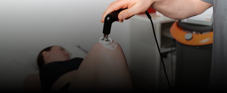
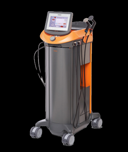
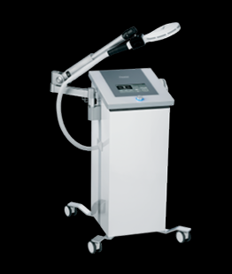
Doctors Kalika and Brosgol are among the first in their field to incorporate orthobiologics to promote tissue healing. Our orthobiologic procedures are guided by high-resolution ultrasound, to ensure proper needle placement and confirm that injected solutions reach their targeted tissues. Treatment results are dramatically enhanced when combined with focused extracorporeal shockwave therapy (fESWT), another area of expertise for Dr. Kalika.
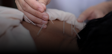
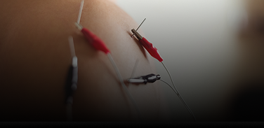
Orthobiologic injection therapies use natural/neutral solutions, injected with precision thanks to ultrasound guidance. The injected solutions stimulate cellular repair by either nourishing or irritating the targeted cells. Needling procedures like dry needling and PENS use filament-thin non-medicated needles to target myofascial trigger points and normalize neural activity.
Knee osteoarthritis is strongly associated with chronic systemic inflammation, prompting doctors to treat the condition with anti-inflammatory drugs and injections. Buit while knee OA involves the degeneration of cartilage, it is considered to be a chronic disease of the entire joint, affecting the cartilage, meniscus, ligaments, and muscles surrounding the knee. Its debilitating nature is strongly associated with metabolic disease and premature death.
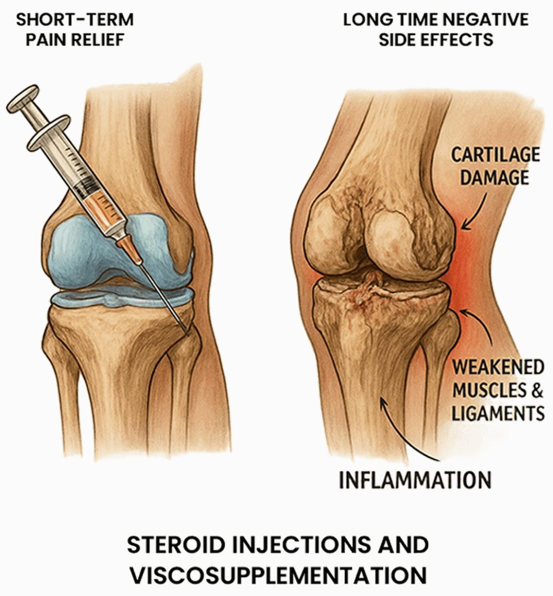
Approaches such as corticosteroid injections to reduce inflammation and viscosupplementation to reduce friction are common standards of care for knee OA. However, the evidence supporting those approaches is losing traction as new research exposes their limitations and adverse side effects that offset any short-term benefits.
A 2021 review of scientific literature found that corticosteroid injections, while moderately effective for short-term pain relief, have multiple adverse side effects ranging from mild to severe.
Steroid negative side effects include:
A 2022 review published in the British Medical Journal explored the effectiveness of viscosupplementation for knee OA. The researchers found that viscosupplementation is not only ineffective compared with placebo – it can have serious adverse side effects and should be used with caution.
When dealing with knee OA, our goal is to help the patient attain the highest level of knee function and mobility. That can only be achieved when the tissues and structures that surround and support the knee are in peak condition.
In our experience, the best outcomes for knee OA require a personalized multimodal approach that leverages the most advanced therapeutic options. By combining orthobiologics, regenerative technologies and manual therapies – along with targeted physical therapy – we get superior and lasting outcomes that restore pain-free functional mobility.
Once your knee tissue has been pretreated and pain and inflammation are under control, we are ready to begin physical therapy. The goal of physical therapy is to restore strength and stability, optimize mobility, and re-establish optimal neuromuscular pathways and muscle coordination patterns.
We don’t just target the knee itself – proper knee function depends on harmonious mobility along the entire kinetic chain. Our advanced interdisciplinary approach allows us to personalize your treatment plan based on your unique patient profile. Many of our patients are able to delay or completely avoid knee replacement surgery.
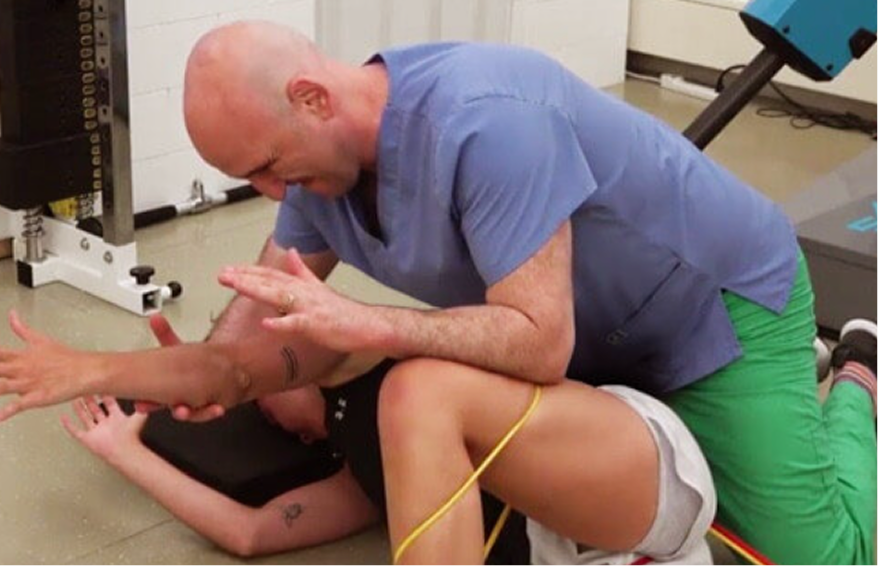
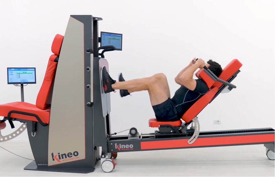
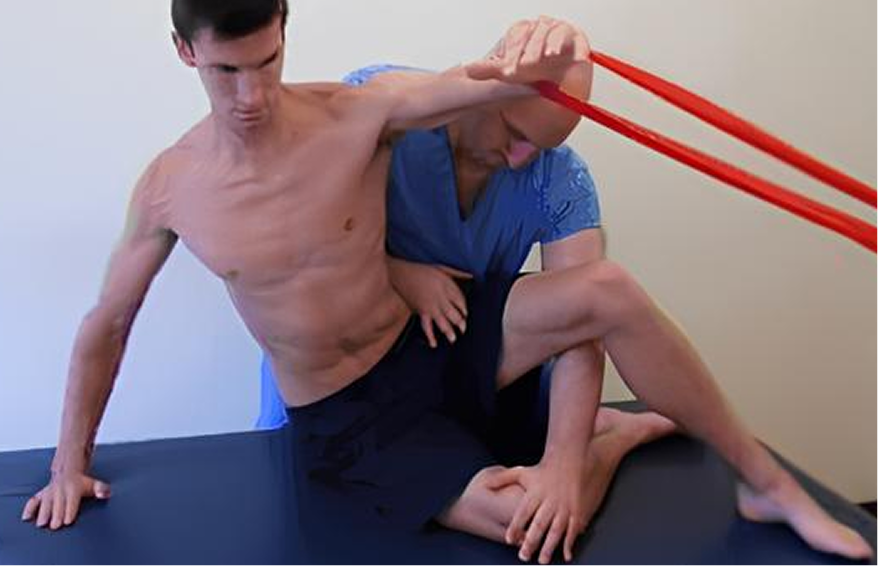
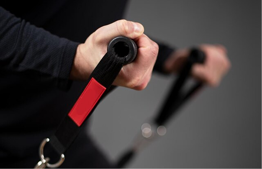
Your personalized physical therapy may include a combination of the following approaches:


Knee osteoarthritis is strongly linked to metabolic health and lifestyle factors. Chronic systemic inflammation can contribute to the development and progression of knee OA due to elevated cytokine levels that cause cartilage degradation, promote synovial inflammation, and alter joint tissue. Obesity, metabolic syndrome, and autoimmune diseases can all increase your risk of knee OA.
There are a number of things you can do to prevent the onset and progression of knee OA:

Medical doctors may try to manage your knee OA symptoms with drugs and surgery without attempting to reverse its progression or improve knee function. In many cases, medical interventions can worsen your condition, leaving you with irreversible knee damage that results in joint replacement surgery.
The clinic at NYDNRehab features the most advanced technologies and cutting-edge therapies for halting the progression of knee OA and reversing cartilage degradation. Our holistic multimodal approach ensures you get the best care available to accelerate healing and restore pain-free knee function. Contact us today, and stop knee OA in its tracks with the most advanced knee OA treatment in NYC.


Clinical director & DC RMSK


 Dr. Yuri Brosgol
MD
Dr. Yuri Brosgol
MD
 Dr. Michael Goynatsky
DPT
Dr. Michael Goynatsky
DPT
 Dr. Daniela Escudero
DPT
Dr. Daniela Escudero
DPT
 Dr. Michelle Agyakwah
DC
Dr. Michelle Agyakwah
DC
 Dr. Tatyana Kapustina
L. Ac.
Dr. Tatyana Kapustina
L. Ac.
Independent peer-reviewed research relevant to this treatment approach.
Research authored or co-authored by the clinic’s medical director. The following research publications inform the clinical approach used in this treatment program.
Conference Abstract
2024
 Lev Kalika
  Rostylav Bubnov
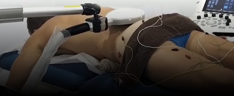
Below is a prime example of how ultrasound can take the guesswork out of diagnosis.

A bad physical therapy experience is one of the primary causes of unnecessary surgery
In this instance, an athlete was originally diagnosed with minor quadriceps muscle strain and was treated for four weeks, with unsatisfactory results. When he came to our clinic, the muscle was not healing, and the patients’ muscle tissue had already begun to atrophy.
Upon examination using MSUS, we discovered that he had a full muscle thickness tear that had been overlooked by his previous provider. To mitigate damage and promote healing, surgery should have been performed immediately after the injury occurred. Because of misdiagnosis and inappropriate treatment, the patient now has permanent damage that cannot be corrected.
The most important advantage of Ultrasound over MRI imaging is its ability to zero in on the symptomatic region and obtain imaging, with active participation and feedback from the patient. Using dynamic MSUS, we can see what happens when patients contract their muscles, something that cannot be done with MRI. From a diagnostic perspective, this interaction is invaluable.
Dynamic ultrasonography examination demonstrating
the full thickness tear and already occurring muscle atrophy
due to misdiagnosis and not referring the patient
to proper diagnostic workup
Demonstration of how very small muscle defect is made and revealed
to be a complete tear with muscle contraction
under diagnostic sonography (not possible with MRI)

Complete tear of rectus femoris
with large hematoma (blood)

Separation of muscle ends due to tear elicited
on dynamic sonography examination