Hip Arthroscopy for FAI syndrome, does it really bring you back to your pre-injury A-game?

Hip Arthroscopy

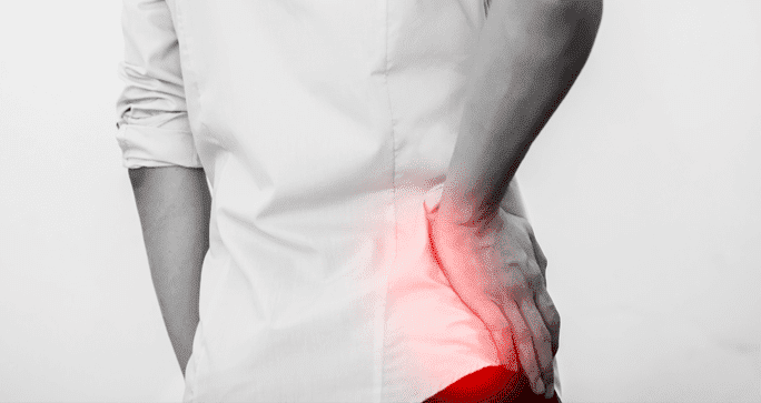
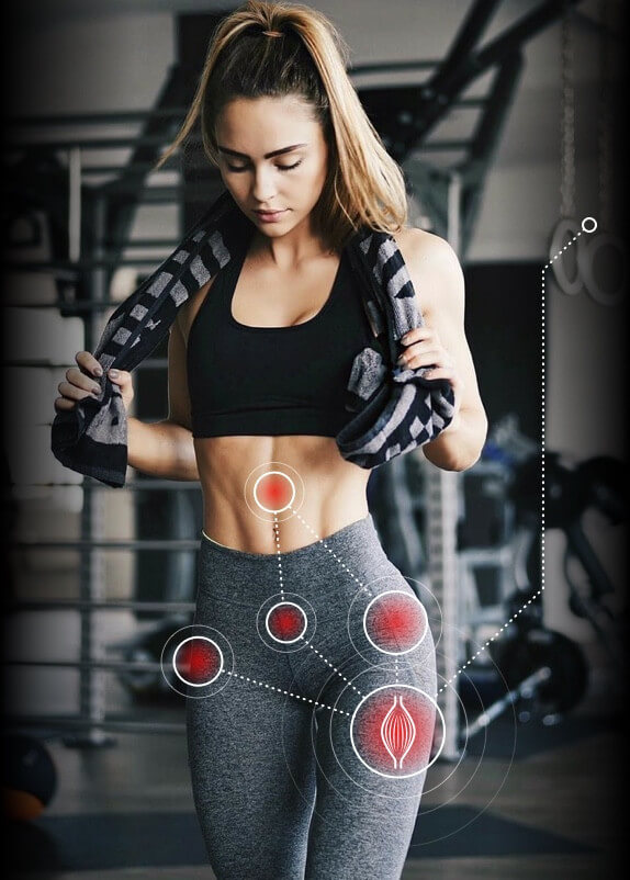
Femoroacetabular impingement syndrome (FAIS) or hip impingement is usually a result of a growth of an extra bone at the hip socket. Since our bones are like a jigsaw puzzle, when this happens the joint no longer works as it should.

Hip impingement can cause extreme pain in the groin. Some of the activities that increase the pain include running, walking jumping and in some cases sitting long periods.

There have been studies aimed at finding out whether having hip arthroscopy helps the situation and gets you back to optimal performance. Though anyone can have an arthroscopy, our analysis will consider athletes. This is because athletes tend to be more active and will be a better measure of the success of the procedure.

Before we begin, we would like to discuss what happens with the studies we have reviewed. However, most of these studies have shown inconsistencies in their results. There are two main reasons for that we are going to get past to get more accurate results.

  1. The first problem is the definition of the result. The expectation is that the patients should go back to doing sports, but the level at which they can do sports is never clearly defined. For this study, we are going to assume that the participants in the survey go back to participating in sports at the level they were before they suffered FAI.
  2. The second issue is the reputation of the practitioner who performs the hip arthroscopy. When all the results for all the procedures are bunched together regardless of where they took place and who did them we aren’t going to have accurate results. We are therefore going for only the procedures in high ranking institutions by renowned physicians.

Who are the participants?

The participants in this analysis are people who have the following characteristic:

  • They are between 18 and 30 years of age
  • They had been participating in sports before they started experiencing hip and groin pain from FAI
  • The persons have gone through a hip arthroscopy and should have gone back to their pre-injury sport.

How did the study work?

The participants were given questionnaires to fill to help with determining how their treatment had gone and if they had the desired results.

In the beginning, there were 350 prospects. But with time the number fell to 189.

Those who did not make it to the end either did not fill in the questionnaire were not into sports before their hip and groin pain, or they had no intention of going back to participating in their pre-injury sports.

The results

When the analysis was done, the participants fell into three categories:

  1. Full sports participation and optimal performance
  2. Full sports participation but less than optimal performance
  3. Less than optimal performance as well as limited participation in sports.

One thing you need to understand was that there was no evidence of the type of sport undertaken and the results. No matter which sports the participants took, they could fall into any category.

Around 29% of the participants fell into category one, 21.4 % were in category two whereas the majority which is 46.4 % had trouble going back to sports and performing well.

Out of those who reported optimal performance, only 8% had been back in the game in within six months and a year of the surgery.

What happens to the group that did not go back to sports?

While there are some who did so to avoid further injury (10%), the majority of them (90%) decided not to go back to active sports because they continually had hip and groin pain.

Final thoughts

The results above show that while hip arthroscopy can help athletes get back to their active lives, only a few of them do and even then, it may only happen after a year in recovery.

Diagnosis

app

A clinical exam and diagnostic ultrasound imaging can help your therapist pinpoint the exact location and cause of your hip and groin pain.

Ultrasound enables you and your therapist to view the hip and groin region in real time, while in motion. In addition to ultrasound, video gait analysis can help us identify faulty movement mechanics that contribute to hip and groin pain. Once the exact cause is determined, an effective treatment plan can be initiated.

img

Explore more advanced diagnostic tools available only at NYDNRehab:

img

Hip dysfunction and pain can be a complex issue due to interactions of the trunk, pelvis, low back, groin and hip joint. Physical therapy and rehabilitation that is based only on subjective clinical analysis often addresses the symptoms without resolving the underlying cause.

icons

At NYDNRehab, our groundbreaking motion analysis technology and high resolution diagnostic ultrasonography have enabled us to develop a battery of tests that perfectly reveal the dynamic functional pathology of the hip joint and pelvis. Our tests are evidence-based protocols that are considered to be the gold standard in the world of research.

Our testing protocol includes:

1

Combined lumbopelvic hip stability test using DLEST methodology with C.A.R.E.N., our computer assisted rehab environment

2

Hip joint stability test using DLEST methodology with C.A.R.E.N.

3

3D star excursion banner test (SEBT) for assessing the involvement of the hip joint and muscles in postural stability

4

3D gait or running analysis

5

3D kinematic joint angle analysis during a squat, lunge, drop jump and pelvis on hip rotation

6

Rehabilitative ultrasonography for viewing intrinsic hip stabilizing muscle activation patterns

We also perform neuromotor testing with DD Robotech for:

  • Proprioception
  • Tracking ability
  • Force sense
  • Critical power
  • Reactive power
  • Flexibility

SEMG

Surface electromyography (SEMG) may be added to any of the above tests when needed.

Based on our experience and evidence-based information, we believe that physical therapy and rehabilitation should be based on objective quantifiable data.

bg

In this instance, an athlete was originally diagnosed with minor quadriceps muscle strain and was treated for four weeks, with unsatisfactory results. When he came to our clinic, the muscle was not healing, and the patients’ muscle tissue had already begun to atrophy.

Upon examination using MSUS, we discovered that he had a full muscle thickness tear that had been overlooked by his previous provider. To mitigate damage and promote healing, surgery should have been performed immediately after the injury occurred. Because of misdiagnosis and inappropriate treatment, the patient now has permanent damage that cannot be corrected.

The most important advantage of Ultrasound over MRI imaging is its ability to zero in on the symptomatic region and obtain imaging, with active participation and feedback from the patient. Using dynamic MSUS, we can see what happens when patients contract their muscles, something that cannot be done with MRI. From a diagnostic perspective, this interaction is invaluable.

Dynamic ultrasonography examination demonstrating
the full thickness tear and already occurring muscle atrophy
due to misdiagnosis and not referring the patient
to proper diagnostic workup

Demonstration of how very small muscle defect is made and revealed
to be a complete tear with muscle contraction
under diagnostic sonography (not possible with MRI)

image

Complete tear of rectus femoris
with large hematoma (blood)

image

Separation of muscle ends due to tear elicited
on dynamic sonography examination

bg
Buy now 3D Gait
Payment Success
Request Telehealth Request Telehealth Request in office visit Book now