Infographic: Is Exercise a Viable Treatment for Plantar Fasciopathy (Chronic Heel Pain)?

-Viable-Treatment-for-Plantar-Fasciopathy

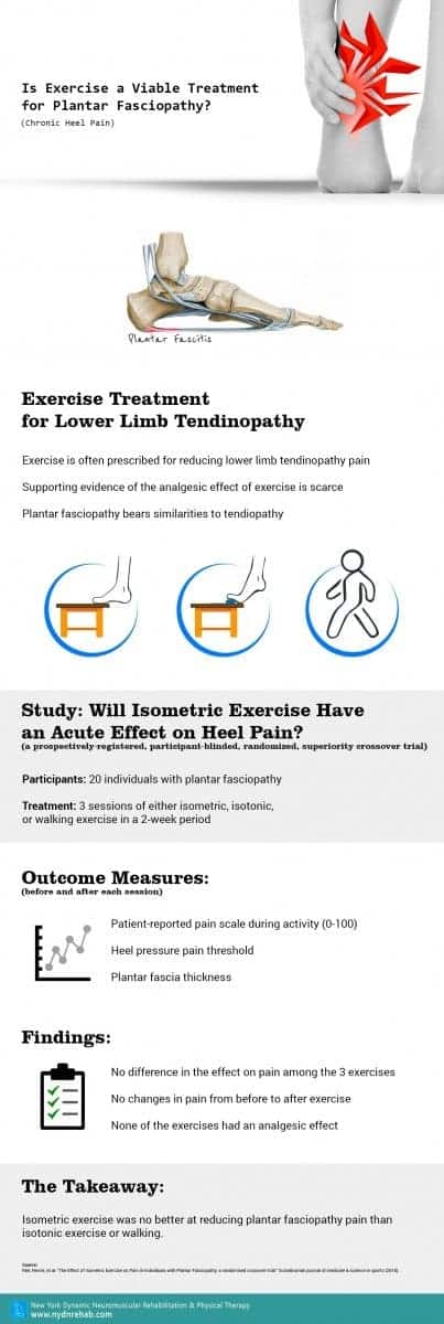
Exercise Treatment for Lower Limb Tendinopathy

  • Exercise is often prescribed for reducing lower limb tendinopathy pain
  • Supporting evidence of the analgesic effect of exercise is scarce
  • Plantar fasciopathy bears similarities to tendiopathy

Study: Will Isometric Exercise Have an Acute Effect on Heel Pain?

(a prospectively‐registered, participant‐blinded, randomized, superiority crossover trial)

  • Participants: 20 individuals with plantar fasciopathy
  • Treatment: 3 sessions of either isometric, isotonic, or walking exercise in a 2‐week period

Outcome Measures (before and after each session):

  • Patient-reported pain scale during activity (0-100)
  • Heel pressure pain threshold
  • Plantar fascia thickness

Findings:

  • No difference in the effect on pain among the 3 exercises
  • No changes in pain from before to after exercise
  • None of the exercises had an analgesic effect

The Takeaway:

Isometric exercise was no better at reducing plantar fasciopathy pain than isotonic exercise or walking.

Chronic-Heel-Pain
bg

In this instance, an athlete was originally diagnosed with minor quadriceps muscle strain and was treated for four weeks, with unsatisfactory results. When he came to our clinic, the muscle was not healing, and the patients’ muscle tissue had already begun to atrophy.

Upon examination using MSUS, we discovered that he had a full muscle thickness tear that had been overlooked by his previous provider. To mitigate damage and promote healing, surgery should have been performed immediately after the injury occurred. Because of misdiagnosis and inappropriate treatment, the patient now has permanent damage that cannot be corrected.

The most important advantage of Ultrasound over MRI imaging is its ability to zero in on the symptomatic region and obtain imaging, with active participation and feedback from the patient. Using dynamic MSUS, we can see what happens when patients contract their muscles, something that cannot be done with MRI. From a diagnostic perspective, this interaction is invaluable.

Dynamic ultrasonography examination demonstrating
the full thickness tear and already occurring muscle atrophy
due to misdiagnosis and not referring the patient
to proper diagnostic workup

Demonstration of how very small muscle defect is made and revealed
to be a complete tear with muscle contraction
under diagnostic sonography (not possible with MRI)

image

Complete tear of rectus femoris
with large hematoma (blood)

image

Separation of muscle ends due to tear elicited
on dynamic sonography examination

bg
Buy now 3D Gait
Payment Success
Request Telehealth Request Telehealth Request in office visit Book now