Chronic knee pain can be debilitating, forcing you to reduce your physical activity level, which in turn can lead to other health issues, including metabolic disease and weight gain. Many people turn to drugs and surgery as a first line of action to reduce knee pain, yet those approaches often fall short of eliminating pain and restoring function.
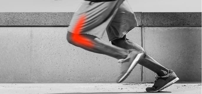
Patellofemoral pain syndrome, sometimes called “runner’s knee” refers to pain in the front of the knee and around the kneecap. The condition is common in athletes in sports that involve running and jumping, and in recreational runners. PFS is caused by overuse and misalignment of the structures of the knee, often due to imbalanced muscle tension or mechanical deficits.
Rehabilitation goals for PFS include
Standard resistance training is a broadly accepted treatment approach for accomplishing those goals. In most cases, loading the quadriceps at 70 percent of one-rep-max (70%/1RM) is effective in strengthening the quads and increasing muscle size (1RM is the maximum load you can lift in one repetition). Most people are able to perform 10 to 12 repetitions at this training load. But for some people, loading at 70%/1RM causes excessive knee pain, ruling it out as a treatment option.
Low-load blood flow restriction (BFR) training is an innovative approach to PFS treatment for patients who cannot tolerate high training loads. The approach uses a blood pressure cuff or band to reduce blood flow to the quadriceps, and training loads are reduced to 30 percent of 1RM. Exercises are performed to volitional fatigue, meaning the exercise is continued until the patient cannot perform another complete repetition.
The mechanisms behind low load BFR training are not entirely clear, yet the approach appears to produce desirable results comparable to traditional resistance training at 70%/1RM.
A recent study by Lachlan et al. (2017) compared the effectiveness of standard resistance training (RT) to low-load BFR training for increasing strength and reducing pain in patients with PFS. Seventy-nine participants with patellofemoral pain were randomly assigned to either standard quadriceps training or low-load BFR. Exercises included one open-chain and one closed-chain exercise (bilateral knee extensions and two-legged leg presses), performed three times per week for eight weeks.
At the end of the training period, 93 percent of the low-load BRF group reported a greater reduction in pain than the standard RT group.
One of the things that sets our clinic apart is our willingness to explore and embrace innovative treatment approaches and technologies, to help our patients eliminate pain and restore function.
During your appointment at NYDNR, your treatment for PFS may include:
You don’t have to live with knee pain. Contact NYDNR today, and see what a difference sports physical therapy can make in improving your performance and overall quality of life.
Source
Giles, Lachlan, et al. “Quadriceps strengthening with and without blood flow restriction in the treatment of patellofemoral pain: a double-blind randomised trial.” Br J Sports Med 51.23 (2017): 1688-1694.

Verified Expert Profiles
Dr. Lev Kalika is a world-recognized expert in musculoskeletal medicine. with 20+ years of clinical experience in diagnostic musculoskeletal ultrasonography, rehabilitative sports medicine and conservative orthopedics. In addition to operating his clinical practice in Manhattan, he regularly publishes peer-reviewed research on ultrasound-guided therapies and procedures. He serves as a peer reviewer for Springer Nature.
Dr. Kalika is an esteemed member of multiple professional organizations, including:
Below is a prime example of how ultrasound can take the guesswork out of diagnosis.

A bad physical therapy experience is one of the primary causes of unnecessary surgery
In this instance, an athlete was originally diagnosed with minor quadriceps muscle strain and was treated for four weeks, with unsatisfactory results. When he came to our clinic, the muscle was not healing, and the patients’ muscle tissue had already begun to atrophy.
Upon examination using MSUS, we discovered that he had a full muscle thickness tear that had been overlooked by his previous provider. To mitigate damage and promote healing, surgery should have been performed immediately after the injury occurred. Because of misdiagnosis and inappropriate treatment, the patient now has permanent damage that cannot be corrected.
The most important advantage of Ultrasound over MRI imaging is its ability to zero in on the symptomatic region and obtain imaging, with active participation and feedback from the patient. Using dynamic MSUS, we can see what happens when patients contract their muscles, something that cannot be done with MRI. From a diagnostic perspective, this interaction is invaluable.
Dynamic ultrasonography examination demonstrating
the full thickness tear and already occurring muscle atrophy
due to misdiagnosis and not referring the patient
to proper diagnostic workup
Demonstration of how very small muscle defect is made and revealed
to be a complete tear with muscle contraction
under diagnostic sonography (not possible with MRI)

Complete tear of rectus femoris
with large hematoma (blood)

Separation of muscle ends due to tear elicited
on dynamic sonography examination


