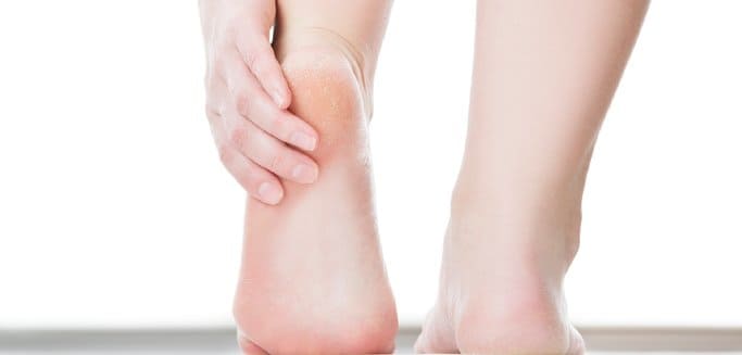
It is really hard to get anything done when your foot is aching so much. It gets really annoying when you cannot even get the most basic tasks of the day done since your foot is in fun let alone run, or do anything that requires loads of footwork. In order to get the pain cured, you do everything possible from resting to put ice on your foot and even foot massages. Still, the pain doesn’t get any better and you fail to identify the underlying reason behind the pain in your foot. Pretty Annoying scenario, isn’t it? Well, you are probably suffering from Plantar Fasciitis.
Since the diagnosis of Plantar Fasciitis is based on the location of the pain, it has become one of the most wrongly analyzed and investigated diseases of the human body. In reality, the presumed cause of the pain, the plantar fascia present in the bottom of the foot, is in most cases not at all injured.
If you don’t use your ankle much, you will start developing pain in the bottom of your foot. This is due to the fact that if you do not use your ankles while exercising or performing the activities of the day, the bottom of your foot will have to work relatively harder. Since it is being overused, the bottom of the foot will start aching after some time.
The reason behind less amount of ankle usage is the development of adhesion in the posterior muscles of the calf. Most of the times, it is the reason behind pain and limited movement in the whole human body. More often than not, correct diagnosis is not made in the cases of Adhesion. Due to overuse and repetitive motion, adhesion can occur in the muscles.
The muscles present at the backside of the lower leg up till the very bottom of the foot are known as the posterior calf muscles. If there is adhesion present in these muscles, you are going to suffer from pain in the bottom of your foot. In order to get a cure for that ache in your foot, the adhesion in the posterior calf muscles needs to be removed.
Now that we have figured out that the major cause behind that foot ache is adhesion, an important question arises. How to make sure if there is adhesion in your muscles? Fortunately, there is an extremely simple test which you can perform at your homes to find it out!
Here is what you need to do:
The most common reason for a limited test is Adhesion. But there can be other reasons as well. You need to find out the reason so that you can get relief from your pain as soon as possible.
With the help of experts at NYDNRehab, you can get the relief from the pain you are looking for! Adhesion is diagnosed and treated in the best way possible at NYDNRehab. Plantar Fasciitis is also treated here in the best way possible.

Verified Expert Profiles
Dr. Lev Kalika is a world-recognized expert in musculoskeletal medicine. with 20+ years of clinical experience in diagnostic musculoskeletal ultrasonography, rehabilitative sports medicine and conservative orthopedics. In addition to operating his clinical practice in Manhattan, he regularly publishes peer-reviewed research on ultrasound-guided therapies and procedures. He serves as a peer reviewer for Springer Nature.
Dr. Kalika is an esteemed member of multiple professional organizations, including:
Below is a prime example of how ultrasound can take the guesswork out of diagnosis.

A bad physical therapy experience is one of the primary causes of unnecessary surgery
In this instance, an athlete was originally diagnosed with minor quadriceps muscle strain and was treated for four weeks, with unsatisfactory results. When he came to our clinic, the muscle was not healing, and the patients’ muscle tissue had already begun to atrophy.
Upon examination using MSUS, we discovered that he had a full muscle thickness tear that had been overlooked by his previous provider. To mitigate damage and promote healing, surgery should have been performed immediately after the injury occurred. Because of misdiagnosis and inappropriate treatment, the patient now has permanent damage that cannot be corrected.
The most important advantage of Ultrasound over MRI imaging is its ability to zero in on the symptomatic region and obtain imaging, with active participation and feedback from the patient. Using dynamic MSUS, we can see what happens when patients contract their muscles, something that cannot be done with MRI. From a diagnostic perspective, this interaction is invaluable.
Dynamic ultrasonography examination demonstrating
the full thickness tear and already occurring muscle atrophy
due to misdiagnosis and not referring the patient
to proper diagnostic workup
Demonstration of how very small muscle defect is made and revealed
to be a complete tear with muscle contraction
under diagnostic sonography (not possible with MRI)

Complete tear of rectus femoris
with large hematoma (blood)

Separation of muscle ends due to tear elicited
on dynamic sonography examination


