Top 12 Tips for Plantar Fasciitis / Heel Pain
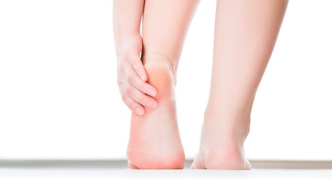
The foot is a truly miraculous structure in which all of the portions of it are connected somehow. Because of this fact, foot injuries are immensely common, specifically that of plantar fasciitis. This affliction is when the plantar fascia tissue that connects the heel to the toes becomes inflamed.
Common indeed, this condition affects over three million people each year and is often self-diagnosed due to the unmistakable pain. Typically, runners are the most affected by this, but truly nobody is exempt from contracting this painful condition at some point in their lives. An acute pain near the heel that is often worse in the morning is the most common symptom of plantar fasciitis, but a plethora of tips exist in favor of combating this painful issue.
Stretch the Fascia
The failure to stretch before any sort of physical activity can be detrimental to the body as a unit, so stretching the fascia can reduce the preexistent heel pain and any other aches in the body.
Simply keeping your heel and arch flat will stretch the toes. Hold this position for ten seconds at least five times a day.
Exercise to Regain Use
Although a professional physical therapist will create an exercise program for the individual and the level of pain that they are experiencing, a common set of exercises exists for battling this heel pain. Of course, the act of exercising will strengthen the rest of the body, thus addressing back, pelvis, leg, and joint pains as well.
Build Strength While Concurrently Stretching the Plantar Fascia
A method known as the High Load has been proven to work effectively. Simply put, this method involves raising the heel while a towel or cloth is under the toes. This action results in regaining mobility back in the foot due to the strength built in this muscle.
Build Leg Strength and Mobility
The human body works together as a unit, so when one portion is afflicted, the other might follow the same path. When the heel is in pain, a person will limp to avoid straining the area. This results in weakened leg muscles, but basic physical therapy can regain mobility and strength in all portions of the body, including the leg.
Roll a Water Bottle Under the Arch
In order to abate the pain of the area, it is recommended to freeze or chill a water bottle.
First, stretch the area and then roll the bottle under the arch. This will promote the mobility of the area while consequently strengthening it.
The Frozen Golf Ball Method
Much like the water bottle activity, freezing a golf ball will serve the same purpose.
However, the ball is firmer, so it massages the area, ultimately reducing the pain.
Elevate the Arch with an Insole
An insole will prevent the plantar fascia from flexing or overexerting itself. This support should be worn at all times and not only during physical activity. This continual action will reduce the effects of plantar fasciitis or prevent it from happening entirely.
Invest in Orthotics
Most of the time, orthotics are sold over the counter and will work well for a multitude of patients. However, certain individuals demonstrate severe plantar fasciitis that has affected other portions of the body, so meeting with a physician to discuss custom orthotics is crucial.
Tape the Fascia
Before investing in orthotics, it is important to attempt something known as Low Dye Taping. Also known as calcaneal taping, this is when the heel portion of the foot is wrapped in a way that will provide it with support and from flexing.
Remain Educated on Plantar Fasciitis
Too often, people attribute this initial pain as being the remnants from a decent workout. The failure to recognize this issue early can pose threatening to the body permanently, so it is best to remain aware of what this condition is, the progressions being made to combat it, and what can be done to protect yourself from ever contracting this issue.
Consider a Night Splint
As previously mentioned, the heel pain associated with this condition is the worst in the morning, but that is often the result of the manner a person slept the night before. A night splint will keep the foot elevated by placing slight pressure on it. This will promote the function of the foot the next morning.
Use Medication Sparingly
If the pain is too much, speak with your physician about medication to alleviate it enough to stretch and gain mobility back to the area.