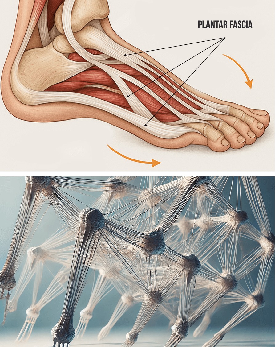
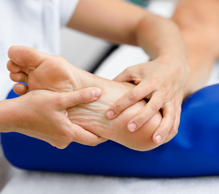
The plantar fascia is a tough band of connective tissue that spans the bottom of the foot from heel to toe, serving to stabilize the many bones that make up your foot and ankle complex. The plantar fascia has elastic properties that store and release energy as it shortens and lengthens, contributing to force production during walking, running and jumping. Although the suffix “itis” is used to describe an inflamed state, it is now evident that plantar fasciitis is not caused by inflammation, but by micro tears and trigger points in the fascia itself.
At NYDNRehab, we are raising the bar for plantar fasciitis treatment by using new regenerative technologies and orthobiologic solutions that accelerate tissue healing and quickly relieve plantar fasciitis pain.

Clinical director & DC RMSK
Verified Expert Profiles
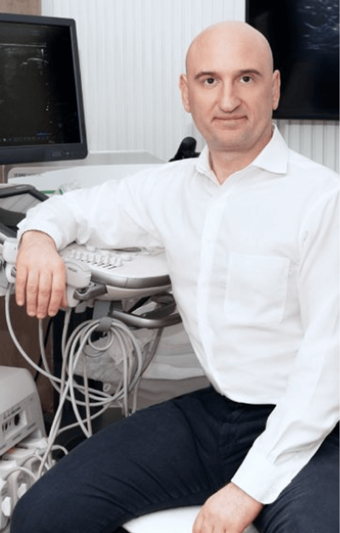
Dr. Lev Kalika, DC, clinical director of NYDNRehab, is an internationally recognized expert in diagnostic and musculoskeletal ultrasonography, with multiple research papers to his credit. Dr. Kalika has studied with some of the world’s most prestigious experts in diagnostic, fascia, and nerve ultrasonography, and has presented his research at multiple international conferences.
Dr. Kalika specializes in the treatment of tendons and fascia, and has pioneered some of the most advanced solutions for dramatically accelerating plantar fasciitis healing. With 20+ years of functional podiatry experience and an impressive track record of success, Dr. Kalika has dedicated his life’s work to advancing holistic therapies that get to the bottom of myofascial pain, without drugs or surgery.
Dr. Kalika is an active member of the American Institute of Ultrasound in Medicine (AIUM), and has developed his own unique approach to Dynamic Functional and Fascial Ultrasonography. He is trained and certified in the Stecco Method of myofascial release.

Orthobiologic, Sports Medicine and Regenerative Medicine Specialist
The plantar fascia is part of a complex network of fascia – tough elastic connective tissue that supports the structure and function of the human body. Fascia works together with muscles, forming the myofascial system that guides and controls movement, and mediates outside forces. Biotensegrity describes the myofascial system’s state of perpetual elastic tension that enables pain-free mobility.
Fascia encases and separates muscles, lines the walls of body cavities, and suspends and supports the internal organs, holding them in place during movement. Healthy fascia is slippery, smooth and elastic, made up of collagenous layers and lubricated by hyaluronan – a fluid composed of hyaluronic acid, able to hold 100X its volume in water. Fascia’s slippery properties enable muscles, tendons, nerves and blood vessels to glide among other structures without friction.
Fascia is densely innervated with mechanoreceptors – sensory nerves that inform your brain of the body’s position in space, and capable of generating pain 5X greater than muscle tissue. When fascia is injured, its layers become dense and sticky, often entrapping nerves and blood vessels, and preventing them from gliding. Proprioception is impaired and biotensegrity is disrupted, causing pain, reducing movement efficiency, and increasing your risk of injury.
Plantar fasciitis is caused by microtears and densified fascial layers that have lost their elastic properties and become sticky. Not only does it cause debilitating heel and foot pain, but a damaged plantar fascia loses its ability to store and release energy – a phenomenon known as the Windlass Mechanism – thus impairing gait and causing mechanical deficiencies throughout the lower kinetic chain.
Most people first see a medical doctor for foot and heel pain, which is frequently misdiagnosed as plantar fasciitis based on the patient’s symptoms. But plantar fasciitis is one of the most commonly misdiagnosed disorders, and multiple other conditions share the same symptoms.
Conditions that mimic plantar fasciitis symptoms include:
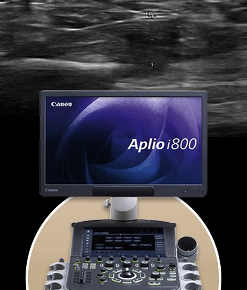
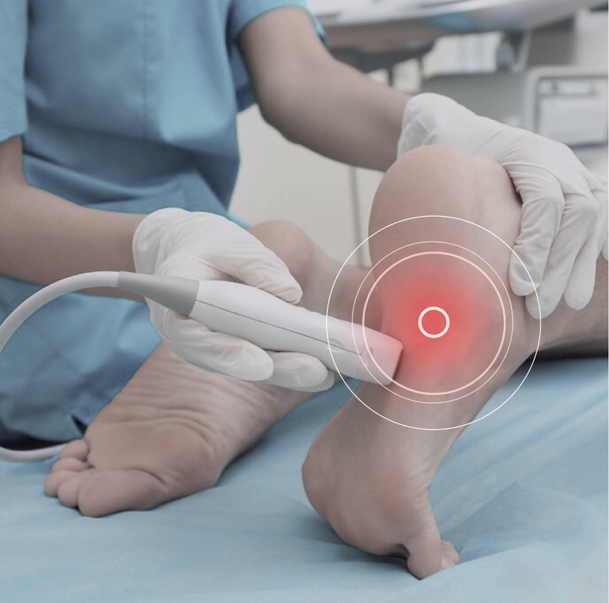
At NYDNRehab, we use high-resolution diagnostic ultrasound to visualize the structures and tissues of the foot and ankle in real time, on your very first visit. Dr. Kalka’s expertise in diagnostic ultrasonography is based on years of training and experience, and an in-depth knowledge of human anatomy. Your in-office ultrasound exam lets us accurately differentiate plantar fasciitis from imposters, so we can quickly proceed with treatment.
Ultrasound imaging gives us a clear picture of what is going on beneath the skin, eliminating guesswork based on symptoms alone, to ensure you get the most effective treatment available. We never take a one-size-fits-all approach to patient care. Our personalized, one-on-one treatment plans are custom-designed to accelerate healing, based on your unique patient profile.
The clinic at NYDNRehab features some of the most advanced technologies and methodologies available in rehabilitative medicine, providing fast and effective pain relief for plantar fasciitis. Patients often report a dramatic reduction in pain after their first treatment session, and complete recovery from plantar fasciitis after just a few sessions.
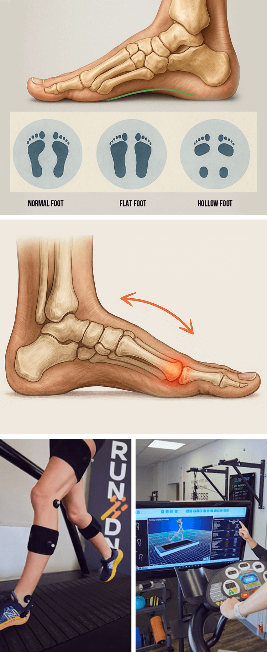
In addition to stress and overuse, plantar fasciitis can arise from biomechanical issues that place excessive stress on the plantar fascia. Conventional treatment zeros in on treating the locus of pain, without addressing other factors that overwork the plantar fascia.
At NYDNRehab, we treat the whole patient, not just your symptoms. Our high-tech motion analysis lab equips us with advanced technologies to quantify movement quality and identify deficiencies that contribute to plantar fascia pain and dysfunction.
Common biomechanical issues that contribute to plantar fasciitis include:
Conventional plantar fasciitis treatment typically involves NSAIDs, corticosteroid injections, and expensive custom orthotics that do little to resolve the condition. Stretching exercises are often prescribed, but they can do more harm than good, especially in the early stages.
Patients often suffer for months on end, going from one doctor to the next in hopes of getting relief. Instead, they are treated with generic, one-size-fits-all protocols that don’t do much good. In many cases, patients don’t have plantar fasciitis at all, but another foot condition that mimics plantar fasciitis symptoms.
Despite being densely populated with mechanoreceptors, the plantar fascia has minimal vascularity. Its limited blood supply is provided primarily via small capillaries that deliver a minimal amount of nutrients and oxygen. Consequently, damaged fascia can be slow to heal, a factor that prolongs plantar fasciitis pain.
In addition to microtears and fascial layer densification, collagen fibers can become disarrayed, and myofascial trigger points can form, interfering with functional foot mechanics. Addressing these issues is the first step toward eliminating pain and restoring function.
At NYDNRehab, our personalized approach means you get a treatment plan tailored to your unique needs. We treat the whole patient, not just your symptoms – our holistic approach focuses on healing and rehabilitation, not just pain management. Our goal is to restore pain-free foot function using cutting-edge technologies and orthobiologic procedures that accelerate the healing process.
At NYDNRehab, we leverage the most recent evidence-based therapies to quickly eliminate plantar fasciitis pain and restore functional mobility. Our multifaceted approach dramatically accelerates the healing process, and patients often enjoy complete recovery after just a few therapy sessions.
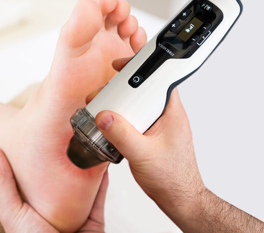
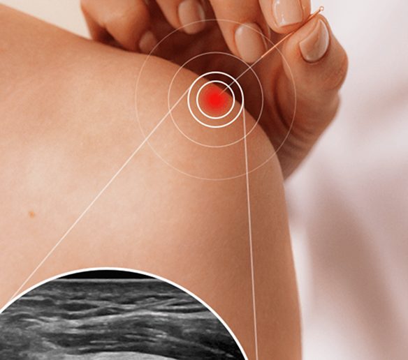
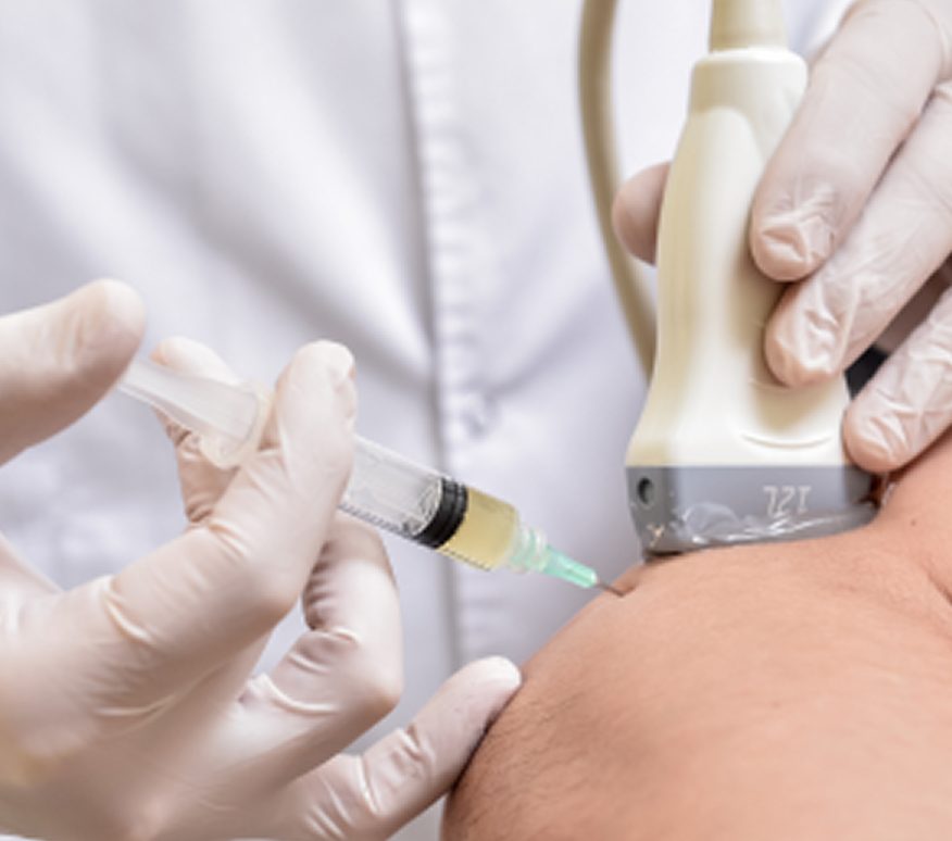
At NYDNRehab, we always begin with a conservative approach to plantar fasciitis treatment, but the secret to our success is precision. We have a 100% track record of success in treating plantar fasciitis, thanks to the combination of extracorporeal shockwave therapy (ESWT) and ultrasound guided dry needling (USDN).
A recent research publication in Nature cites Dr. Kalika’s own research, highlighting USDN as a top-tier evidence-based solution for plantar fasciopathy, and ranking it alongside ESWT and PRP as a primary intervention. To quote the article:
“Current therapeutic approaches for plantar fasciopathy include targeted mechanotherapy, optimized ESWT protocols, ultrasound-guided delivery of orthobiologics (e.g. leukocyte-rich PRP), and precision physical therapies. Furthermore, studies demonstrate the diagnostic and therapeutic value of ultrasound elastography and US-guided interventions for pain modulation. Ultrasound-guided dry needling of myofascial trigger points has been established as a primary intervention option for plantar fasciopathy.”
For patients, our multimodal approach to plantar fasciitis means quick pain relief, fast recovery, and longer-lasting outcomes. In our experience, combining modalities with precision guidance creates the best results.
Plantar fasciitis is a common complaint, especially among people who spend a lot of time on their feet. But you can avoid plantar fasciitis by adopting these preventative strategies:

Plantar fasciitis can be slow to heal, causing pain and reducing mobility for months on end. Conventional therapies like NSAIDs, bracing and orthotics have little evidence to support their efficacy. At NYDNRehab, our holistic evidence-based approach ensures that our patients get the most current and effective therapy possible, to dramatically accelerate recovery from plantar fasciitis and restore pain-free functional mobility. Our expertise, experience and track record of success make NYDNRehab the clinic of choice for plantar fasciitis rehab in NYC.


Independent peer-reviewed research relevant to this treatment approach.

Below is a prime example of how ultrasound can take the guesswork out of diagnosis.

A bad physical therapy experience is one of the primary causes of unnecessary surgery
In this instance, an athlete was originally diagnosed with minor quadriceps muscle strain and was treated for four weeks, with unsatisfactory results. When he came to our clinic, the muscle was not healing, and the patients’ muscle tissue had already begun to atrophy.
Upon examination using MSUS, we discovered that he had a full muscle thickness tear that had been overlooked by his previous provider. To mitigate damage and promote healing, surgery should have been performed immediately after the injury occurred. Because of misdiagnosis and inappropriate treatment, the patient now has permanent damage that cannot be corrected.
The most important advantage of Ultrasound over MRI imaging is its ability to zero in on the symptomatic region and obtain imaging, with active participation and feedback from the patient. Using dynamic MSUS, we can see what happens when patients contract their muscles, something that cannot be done with MRI. From a diagnostic perspective, this interaction is invaluable.
Dynamic ultrasonography examination demonstrating
the full thickness tear and already occurring muscle atrophy
due to misdiagnosis and not referring the patient
to proper diagnostic workup
Demonstration of how very small muscle defect is made and revealed
to be a complete tear with muscle contraction
under diagnostic sonography (not possible with MRI)

Complete tear of rectus femoris
with large hematoma (blood)

Separation of muscle ends due to tear elicited
on dynamic sonography examination


