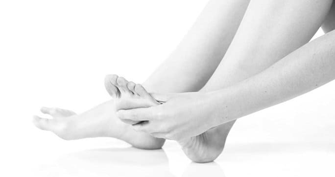
Tired aching dogs can make you miserable and keep you from doing the things you love. Yet many people live with foot pain, accepting it as a normal part of aging. However most foot pain emanates from a specific cause that can often be identified and corrected.
Many daily activities contribute to foot pain, including standing for long hours on concrete, wearing fashion footwear, exercising in worn or inappropriate athletic shoes, wearing the wrong shoe size, and many more. Foot pain can also be caused by certain categories of disease, and by deficient movement mechanics during walking or running.
Whether your foot pain originates from without or within, ignoring it can lead to serious problems that can become debilitating over time.
Foot Structure and Function
Together, your foot and ankle form a complex system made up of 28 bones, 33 joints, and 112 ligaments, all controlled by 13 extrinsic and 21 intrinsic muscles. Their complexity allows for a range of important functions, including:
balance
shock absorption
support of body weight
transfer of ground reaction forces
force production for walking, running and jumping
The foot can be divided into three segments: rearfoot, midfoot, and forefoot. The foot’s unique structure allows for rigidity when standing, and flexibility when moving over uneven terrain.
The Gait Cycle
The gait cycle sums up the series of movements that take place during human walking or running. A complete gait cycle begins when one foot makes contact with the ground and ends when that same foot makes contact with the ground again.
The gait cycle can be broken down into two phases:
Stance Phase, when some part of the foot is in contact with the ground. The stance phase can further be broken down into heel strike, foot flat, mid-stance, heel-off, and toe-off.
Swing Phase, when the foot is traveling through the air as the leg swings forward to initiate the next step.
Since your foot bears your body weight and absorbs impact during the stance phase, it is most important when assessing gait mechanics relating to foot pain.
One way to tell if your foot pain is caused from inefficient movement mechanics is to have a computerized gait analysis. Faulty gait mechanics can lead to a plethora of injuries, not only to the foot, but to the ankle, knee, hip, pelvis and spine.
Poor gait mechanics can and should be corrected to reduce your risk of injury and improve walking and running performance.
Common Foot Injuries
With the complexity of structures that make up your foot, it should not come as a surprise that there are numerous types of foot injuries that can occur. Among the most common are:
Plantar Fasciitis: A common and chronic condition that develops over time, plantar fasciitis usually begins as heel pain that spreads and becomes progressively worse if left untreated.
Navicular Stress Fracture: This is the most common stress fracture to the navicular bone in the midfoot.
Lisfranc Injury: Also occurring in the midfoot, this injury is a dislocation or fracture of the first tarsometatarsal joint.
Metatarsalgia: Occurring at the ball of the foot, this painful condition is caused by an over-prominent metatarsal head.
Hallux valgus: Commonly known as a bunion, the big toe becomes deviated, resulting in foot deformity.
Hallux limitus: Also occurring at the big toe, this condition is marked by limited range of motion at the first metatarsal phalangeal joint.
In addition to injuries, diseases like peripheral neuropathy associated with diabetes, and arthritis associated with age and chronic inflammation, can also cause foot pain. Feet with high arches and flat feet often experience pain.
Diagnosis and Treatment of Foot Pain
Poor mechanics, inappropriate footwear, repetitive stress and compensatory movement patterns are often at the root of foot pain and injury. All of these catalysts can be identified and corrected. A thorough analysis by a trained foot pain specialist is the first step in getting to the source of your foot pain.
Failure to seek treatment for your foot pain may lead to further injury and eventual disability. A dysfunctional foot can also affect your balance, making you prone to injuries from falls.
Treatment for foot pain may include:
Diagnostic ultrasonography
Video gait analysis and retraining
Strengthening and stretching exercises
Stability and balance exercises
ESWT (extracorporeal shockwave therapy)
Cold and heat therapy
At NYDNRehab, our foot pain specialists conduct a thorough analysis to identify and treat the source of your foot pain. We especially focus on movement mechanics, correcting deficient running and walking gait patterns so the foot and ankle move in more mechanically efficient ways.
The foot pain specialists at NYDNRehab go beyond just treating your pain by correcting the underlying deficiencies that often lead to injury. Do not ignore your ongoing foot pain. Contact NYDNRehab today for complete analysis, diagnosis and state-of-the-art treatment.
In this instance, an athlete was originally diagnosed with minor quadriceps muscle strain and was treated for four weeks, with unsatisfactory results. When he came to our clinic, the muscle was not healing, and the patients’ muscle tissue had already begun to atrophy.
Upon examination using MSUS, we discovered that he had a full muscle thickness tear that had been overlooked by his previous provider. To mitigate damage and promote healing, surgery should have been performed immediately after the injury occurred. Because of misdiagnosis and inappropriate treatment, the patient now has permanent damage that cannot be corrected.
The most important advantage of Ultrasound over MRI imaging is its ability to zero in on the symptomatic region and obtain imaging, with active participation and feedback from the patient. Using dynamic MSUS, we can see what happens when patients contract their muscles, something that cannot be done with MRI. From a diagnostic perspective, this interaction is invaluable.
Dynamic ultrasonography examination demonstrating the full thickness tear and already occurring muscle atrophy due to misdiagnosis and not referring the patient to proper diagnostic workup
Demonstration of how very small muscle defect is made and revealed to be a complete tear with muscle contraction under diagnostic sonography (not possible with MRI)
Complete tear of rectus femoris with large hematoma (blood)
Separation of muscle ends due to tear elicited on dynamic sonography examination