New York Dynamic Neuromuscular Rehabilitation & Physical Therapy
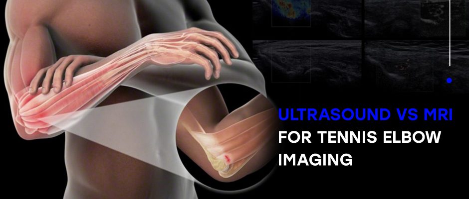
Chronic lateral epicondylitis — aka tennis elbow – is a painful condition caused by repetitive overuse of the forearm wrist extensor muscles that attach to the epicondyle of the humerus bone, just above your elbow. Although tennis elbow can occur at any age, the condition is most commonly reported in adults aged 35-60. Despite its […]
Read More
Hamstring strains are common in sports like football, soccer and other sports that demand high speed kicking, rapid directional changes, and quick deceleration. Dancers are also prone to hamstring strains, with 34 percent reporting at least a single incident. Once an athlete injures a hamstring, re-injury risk goes up, causing the athlete to play. The […]
Read More
NYDNRehab is pleased to announce our 2017 scholarship winners, Greg Stavinski and Daniel Kuchter. Congratulations! Scholarship applicants were required to demonstrate a strong interest in biomedical related research, and an understanding of biomechanics and movement science as applies to rehabilitation. They were also required to articulate their perspectives on technology’s role in the future of […]
Read More
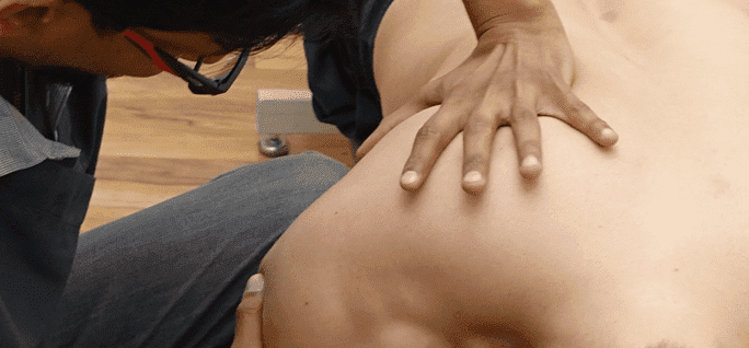
In most cases muscle knots, often called myofascial trigger points, are nothing more than a mere annoyance, causing mild to moderate pain when pressure is applied. However, depending on their location, muscle knots can cause referred pain in other areas of your body that is easy to misdiagnose. What exactly are muscle knots? The cells […]
Read More
Tendons are thick fibrous tissue that attach muscles tor-cuff-tendinitis/” target=”_blank”>Tendinitis pain is most commonly experienced near ankles, knees, wrists, elbows and shoulders. If you experience pain and inflammation near your joint that does not diminish after a few days, contact NYDNRehab for diagnosis and treatment. The sports medicine team at NYDNRehab combines expertise and experience […]
Read More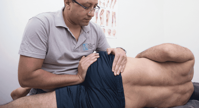
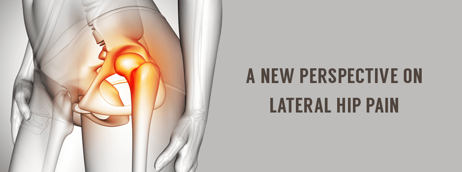
Hip pain can be both distracting and debilitating, nagging at you with each step or movement. While hip pain is sometimes indicative of a structural problem within the joint (intra-articular), pain can often stem from other sources outside of the joint (extra-articular). Some common causes of extra-articular hip joint pain include: ● Tendon injuries […]
Read More
Below is a prime example of how ultrasound can take the guesswork out of diagnosis.

A bad physical therapy experience is one of the primary causes of unnecessary surgery
In this instance, an athlete was originally diagnosed with minor quadriceps muscle strain and was treated for four weeks, with unsatisfactory results. When he came to our clinic, the muscle was not healing, and the patients’ muscle tissue had already begun to atrophy.
Upon examination using MSUS, we discovered that he had a full muscle thickness tear that had been overlooked by his previous provider. To mitigate damage and promote healing, surgery should have been performed immediately after the injury occurred. Because of misdiagnosis and inappropriate treatment, the patient now has permanent damage that cannot be corrected.
The most important advantage of Ultrasound over MRI imaging is its ability to zero in on the symptomatic region and obtain imaging, with active participation and feedback from the patient. Using dynamic MSUS, we can see what happens when patients contract their muscles, something that cannot be done with MRI. From a diagnostic perspective, this interaction is invaluable.
Dynamic ultrasonography examination demonstrating
the full thickness tear and already occurring muscle atrophy
due to misdiagnosis and not referring the patient
to proper diagnostic workup
Demonstration of how very small muscle defect is made and revealed
to be a complete tear with muscle contraction
under diagnostic sonography (not possible with MRI)

Complete tear of rectus femoris
with large hematoma (blood)

Separation of muscle ends due to tear elicited
on dynamic sonography examination