The elbow is a complex hinge joint that assists in arm flexion and wrist rotation. However, it is also especially susceptible to tendinitis, sprains, strains, and medial and lateral epicondylitis. Fortunately, physician-approved programs of exercise for elbow pain and elbow pain support can help the injured patient recover strength and range of motion in the […]
Read MoreOsteoarthritis and bursitis are two musculoskeletal conditions that can afflict the hips. Hip osteoarthritis is a degenerative condition affecting the joints of the hips, while hip bursitis is an inflammation of the bursa at the lateral point of the greater trochanter (the bony point on the side of the body that we normally associate with […]
Read MoreShockwave treatment for tennis elbow is a relatively new form of therapy in which gentle pressure waves are released into the skin around the lateral epicondyle, a bony protrusion of the outer elbow. When used in combination with conventional treatments such as elbow supports and pain relief exercises, shockwave treatment for tennis elbow has proven […]
Read More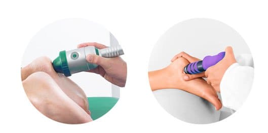
When compared, Extracorporeal shockwave therapy (ESWT) and radial shockwave therapy (RSWT) have many differences. How they are used is the main difference, the type of energy along with it’s density as well as the depths into the muscle tissue. The end result of the stimulation and the mechanism of these two therapies are similar. ESWT […]
Read MorePinched nerve treatment is treatment for nerves that have been entrapped or in some other way compromised. A nerve root is the segment of a nerve leaving the spinal column, and when this nerve becomes compressed, a patient may suffer from severe pain and inflammation. At New York Dynamic Rehabilitation clinic (NYDNRehab), we have years […]
Read MoreHerniation refers together, the joints that allow the spine what mobility it has. When a disc is herniated, or “slipped,” or “ruptured,” the jelly-like center of the disc has begun pushing outwards from within. This can occur because of a sudden rupture or through the natural wear and tear of aging. Because the spinal structure […]
Read MoreThe Achilles tendon is the largest tendon in the human body and runs from a person’s heelbone to all manner of injuries, including chronic Achilles tendinopathy and Achilles insertional tendinopathy. Classical Achilles tendinopathy is the swelling and inflammation of the Achilles tendon. Achilles insertional tendinopathy is the degeneration of the tendon right where it attaches to a complete rupture. The […]
Read MoreWe are all somewhat familiar with how physical therapy can help people with various developmental and physical problems. However, with so much information available top 10 things you might not be aware about physical therapy. Physical therapists do much more than just treating injuries they can help you avoid repetitive injuries like joint and muscle […]
Read More
Below is a prime example of how ultrasound can take the guesswork out of diagnosis.

A bad physical therapy experience is one of the primary causes of unnecessary surgery
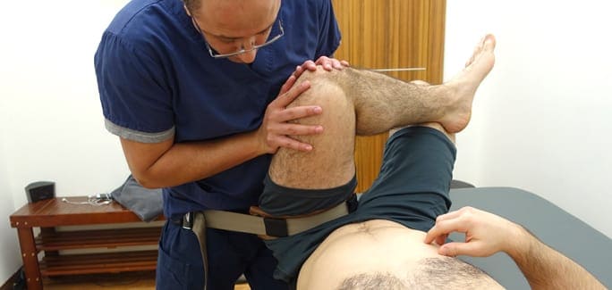
In this instance, an athlete was originally diagnosed with minor quadriceps muscle strain and was treated for four weeks, with unsatisfactory results. When he came to our clinic, the muscle was not healing, and the patients’ muscle tissue had already begun to atrophy.
Upon examination using MSUS, we discovered that he had a full muscle thickness tear that had been overlooked by his previous provider. To mitigate damage and promote healing, surgery should have been performed immediately after the injury occurred. Because of misdiagnosis and inappropriate treatment, the patient now has permanent damage that cannot be corrected.
The most important advantage of Ultrasound over MRI imaging is its ability to zero in on the symptomatic region and obtain imaging, with active participation and feedback from the patient. Using dynamic MSUS, we can see what happens when patients contract their muscles, something that cannot be done with MRI. From a diagnostic perspective, this interaction is invaluable.
Dynamic ultrasonography examination demonstrating
the full thickness tear and already occurring muscle atrophy
due to misdiagnosis and not referring the patient
to proper diagnostic workup
Demonstration of how very small muscle defect is made and revealed
to be a complete tear with muscle contraction
under diagnostic sonography (not possible with MRI)

Complete tear of rectus femoris
with large hematoma (blood)

Separation of muscle ends due to tear elicited
on dynamic sonography examination