
In the human body, fascia is a type of connective tissue made up of thin films of tissue that surround and encase skeletal muscle fibers. Fascia promotes smooth movement by enabling one muscle or muscle fiber to move independently from neighboring tissues. Deep fascia in the back and limbs is made of dense sheets of connective tissue with large numbers of collagen fibers. Deep fascia serves to contain and separate groups of muscles into well-defined spaces called compartments, integrating them and transmitting loads between them.
The dominant cells in fascia are fibroblasts — cells that are able to communicate with each other through gap junctions, and respond to shape changes when tissue is stretched. During wound healing, fibroblasts take on the characteristics of smooth muscle cells, becoming myofibroblasts, with contractile properties that help close a wound. In areas where ligaments, tendons and joint capsules attach to bony structures, connective tissue often flares out to better grip the bone, with fascia linking those tissues to neighboring structures.
When fascia becomes damaged or dysfunctional due to overuse or injury, it can have a profound impact on muscle function. Myofascial decompression therapy is designed to heal and rejuvenate muscle fascia, promoting improved muscle function and performance.
Uses of myofascial cupping therapy include:
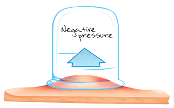
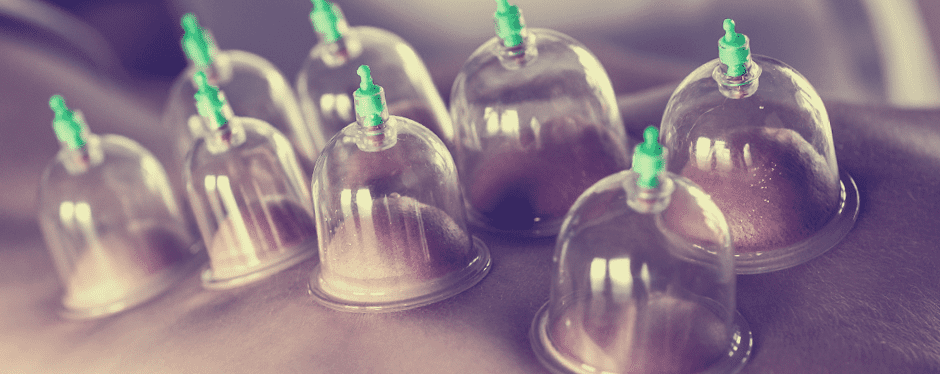
During a cupping session, your therapist will place round glass or plastic domes over the affected area. Using a hand pump, air is suctioned out, creating a vacuum. The vacuum draws the skin and soft tissues upward, creating a negative pressure that is the opposite of massage therapy. As superficial tissues are lifted, deep facia tissues receive more blood, reducing stiffness and promoting healing.
Cupping is able to stimulate acupressure points, creating a similar effect to acupuncture. Bruising is an inherent side effect of cupping therapy, leaving dark purplish-red marks that gradually fade over the course of several days or weeks.
The physical therapy team at NYDNR has advanced training in myofascial decompression techniques. Athletes, physically active patients, and patients who have undergone certain types of trauma or surgery can all benefit from cupping therapy.
Myofascial decompression is particularly effective for several conditions, including:
Cupping therapy should be followed up by neuromuscular re-education, to restore functional movement patterns that were formerly constricted by damaged fascial tissue. DNS (dynamic neuromuscular stabilization) therapy may be useful in the re-education process.

Verified Expert Profiles
Dr. Lev Kalika is a world-recognized expert in musculoskeletal medicine. with 20+ years of clinical experience in diagnostic musculoskeletal ultrasonography, rehabilitative sports medicine and conservative orthopedics. In addition to operating his clinical practice in Manhattan, he regularly publishes peer-reviewed research on ultrasound-guided therapies and procedures. He serves as a peer reviewer for Springer Nature.
Dr. Kalika is an esteemed member of multiple professional organizations, including:
Below is a prime example of how ultrasound can take the guesswork out of diagnosis.

A bad physical therapy experience is one of the primary causes of unnecessary surgery
In this instance, an athlete was originally diagnosed with minor quadriceps muscle strain and was treated for four weeks, with unsatisfactory results. When he came to our clinic, the muscle was not healing, and the patients’ muscle tissue had already begun to atrophy.
Upon examination using MSUS, we discovered that he had a full muscle thickness tear that had been overlooked by his previous provider. To mitigate damage and promote healing, surgery should have been performed immediately after the injury occurred. Because of misdiagnosis and inappropriate treatment, the patient now has permanent damage that cannot be corrected.
The most important advantage of Ultrasound over MRI imaging is its ability to zero in on the symptomatic region and obtain imaging, with active participation and feedback from the patient. Using dynamic MSUS, we can see what happens when patients contract their muscles, something that cannot be done with MRI. From a diagnostic perspective, this interaction is invaluable.
Dynamic ultrasonography examination demonstrating
the full thickness tear and already occurring muscle atrophy
due to misdiagnosis and not referring the patient
to proper diagnostic workup
Demonstration of how very small muscle defect is made and revealed
to be a complete tear with muscle contraction
under diagnostic sonography (not possible with MRI)

Complete tear of rectus femoris
with large hematoma (blood)

Separation of muscle ends due to tear elicited
on dynamic sonography examination


