New York Dynamic Neuromuscular Rehabilitation & Physical Therapy
More and more medical experts recommend chiropractic treatment. They recognize that numerous clinical trials have confirmed the efficacy of this method. Research also proves that chiropractors offer competitive pricing and satisfy patients without compromising safety. Long-Term Pain A British study looked at the impact of chiropractic therapy on individuals who experienced chronic pain in their […]
Read More
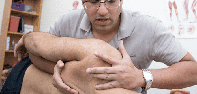
The pinched nerve theory suggests that muscular or bony obstructions catching a nerve cause pain during movement in the affected area. It also suggests that by removing those obstructions, the pinched nerve would be freed if the condition became a perpetual issue. Today, many top neurophysiologists agree that this is an outdated theory. They suggest […]
Read More
Hamstring strains are common sports ailments. Many times, problems occur after previous strains. It is important to learn how Nordic hamstring exercises can prevent and help in the healing of these types of strains. Reasons to Perform Nordic Hamstring Exercises The best way to a solid routine, a person can improve movement and function and […]
Read More
A disorder affecting the elbows and causing 4 percent of cases involving suspected lateral epicondylalgia, radial tunnel syndrome, or RTS, is a painful condition that athletes experience more frequently than others. Correctly assessing and diagnosing RTS is particularly challenging because there are several alternative diagnoses that clinicians must rule out. After it has been diagnosed, […]
Read More
Many people wrongly assume that only those who have been injured or have severe enough problems for a docto get in shape but do not always develop a healthy routine. Pinpointing The Problem In March, it is common to the gym. Some people sto strengthen are not showing improvement. How A Physiotherapist Can Help Physiotherapists […]
Read More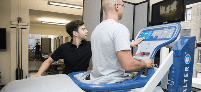
Would you like to run long distances without the harmful effects of pounding your feet against a hard surface? You can accomplish this by using an anti-gravity treadmill. This equipment features sophisticated NASA technology that minimizes the negative aspects of running and promotes rapid rehabilitation. Advantages The treadmill lets you run in a completely natural […]
Read More
Clinical research gives us the best possible information about how best to treat individual patients. What Research Is Appropriate for Evidence-Based Medicine? Evidence-based medicine requires published, peer-reviewed research that doctors of treatment plans can evaluate and verify. Ideally, the authors of the research should support their findings with randomized clinical trials. Trials generally provide ample […]
Read More
When an athlete’s hamstring becomes injured, the individual will experience tightness that significantly restricts the person’s movements and severe pain that occurs while the muscles contract. The person may choose a rehabilitation program that requires eccentric training, or the athlete can participate in a program that strengthens the muscles of the core and enhances the […]
Read More
Below is a prime example of how ultrasound can take the guesswork out of diagnosis.

A bad physical therapy experience is one of the primary causes of unnecessary surgery
In this instance, an athlete was originally diagnosed with minor quadriceps muscle strain and was treated for four weeks, with unsatisfactory results. When he came to our clinic, the muscle was not healing, and the patients’ muscle tissue had already begun to atrophy.
Upon examination using MSUS, we discovered that he had a full muscle thickness tear that had been overlooked by his previous provider. To mitigate damage and promote healing, surgery should have been performed immediately after the injury occurred. Because of misdiagnosis and inappropriate treatment, the patient now has permanent damage that cannot be corrected.
The most important advantage of Ultrasound over MRI imaging is its ability to zero in on the symptomatic region and obtain imaging, with active participation and feedback from the patient. Using dynamic MSUS, we can see what happens when patients contract their muscles, something that cannot be done with MRI. From a diagnostic perspective, this interaction is invaluable.
Dynamic ultrasonography examination demonstrating
the full thickness tear and already occurring muscle atrophy
due to misdiagnosis and not referring the patient
to proper diagnostic workup
Demonstration of how very small muscle defect is made and revealed
to be a complete tear with muscle contraction
under diagnostic sonography (not possible with MRI)

Complete tear of rectus femoris
with large hematoma (blood)

Separation of muscle ends due to tear elicited
on dynamic sonography examination


